By Kris Dickie, Vice President for Research and Development for Clarius.
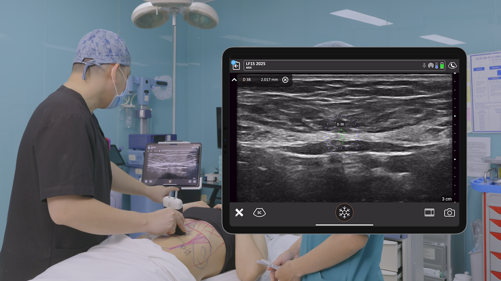
Over the years, ultrasound has become popular for helping to more accurately guide procedures such as nerve blocks, PRP injections, and other routine needle insertions.
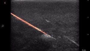
Seeing the needle in tissue on ultrasound has always been a problem that ultrasound manufacturers have striven to overcome. Many high-end system providers introduced a new technique some years ago using beam steering to enhance the needle to see it more clearly in tissue.
Now, beam steering has become standard in many point-of-care ultrasound systems, but the most common technique requires user intervention. Clarius has developed a technique that helps users visualize the needle more effectively, while reducing potential complications and procedure time.
Some technical detail
On most systems, the user needs to manually adjust the beam steer angle, and then the system overlays a processed and high-contrast filtered angled image over the regular B mode to make the needle ‘magically ‘ appear. This method works well; however it forces user intervention depending on which side of the transducer the needle is inserted from, and adjustment when the needle insertion angle changes. Performing this adjustment while using one hand to scan and one hand to insert the needle, is not always simple, since we don’t have a third hand.
Clarius uses a new patent-pending needle enhancement technology that uses a multi-angle approach that adds two unique techniques for needle visualization. First, instead of having the user adjust the angle to ensure perpendicularity to the needle, multiple angles are imaged on both sides of the Scanner. This technique removes user intervention, however, depending on the tissue, imaging artifacts from other fascia or bone may resonate within the other angles showing bright lines that are not useful or diagnostic to the user.
The second technique finds the actual needle, and does more than just apply a high-contrast filter. By looking at metallic needle signatures that technologies like machine-learning algorithms can learn to identify, potential anatomical artifacts are completely removed, and only the true needle will be shown, and thereby tracked through image processing. With the location of the needle always known, angles can then be adjusted in real-time, as can overlay colors and trajectory predictions.
Our needle enhancement technique is another step forward in our ongoing pursuit to simplify ultrasound, while bringing high-end imaging and features in an affordable ultrasound package.
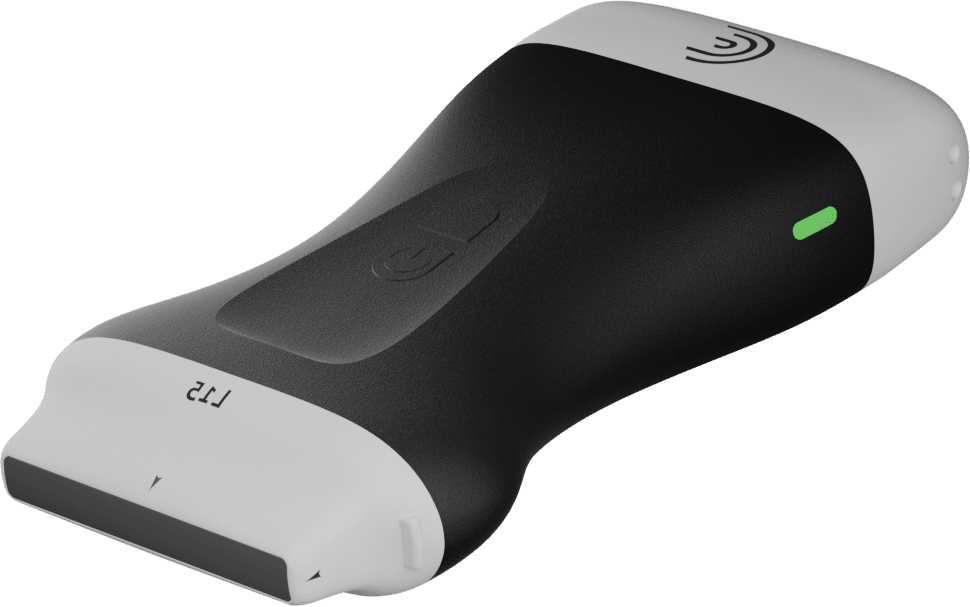
Needle Enhance is an option that is available now with the Clarius L7 Scanner in regions where we have regulatory clearance.
