


The ability to see beneath the skin’s surface non-invasively and in real time, has profoundly transformed the way aesthetic treatments are planned, designed, and performed. Facial ultrasound has emerged as an invaluable tool for clinicians in recent years.
Dr. Karina Ravera is one of the leading experts in the field of ultrasound-guided facial treatments. An aesthetic radiologist and educator based in Argentina, she brings more than 25 years of experience to her field. Dr. Ravera is also the medical director of UltraSkinUS and director of Derma Academy in Latin America. During a recent one-hour Clarius webinar, Dr. Ravera discussed how high-resolution ultrasound, specifically the Clarius L20 HD3, can help clinicians achieve safer, more precise, and more effective outcomes. The webinar is available now to watch at your convenience. Read on for key takeaways.
The Importance of High-Resolution Ultrasound for Facial Anatomy
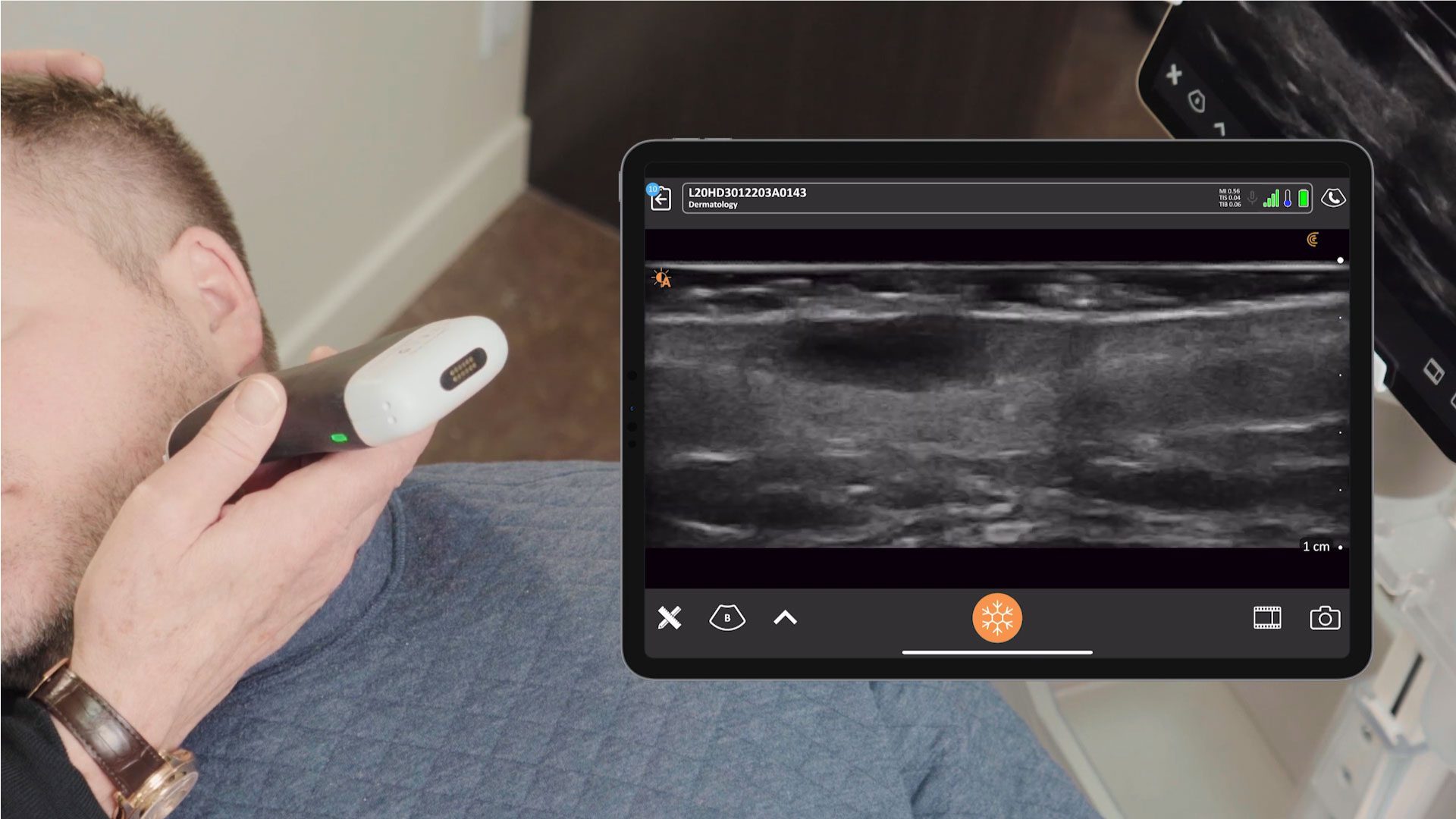
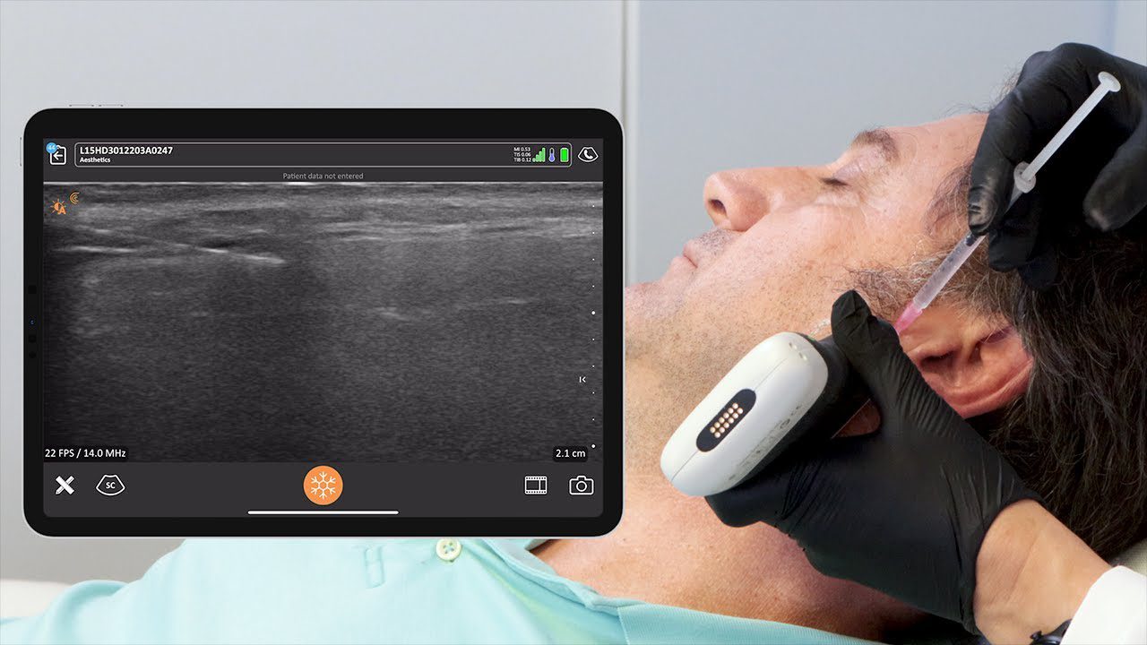
High-resolution ultrasound is the imaging technique that most closely resembles histologic images of the skin, making it the «gold standard for soft tissue assessment,» says Dr. Ravera. By using a linear probe like the Clarius L20 or L15 and applying a soft touch to avoid distorting tissues, clinicians can identify crucial facial structures and layers. This includes the epidermis, dermis, hypodermis, muscles, and bony surfaces.
Key Clinical Takeaways
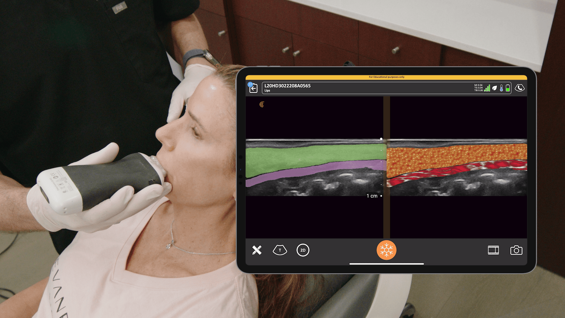
Skin Layer Assessment: The skin is composed of three layers: the hyperechoic epidermis (outer layer), the dermis (intermediate layer rich in collagen and elastin), and the hypodermis (deep layer containing fat globules). These layers appear differently on an ultrasound, with the hypodermis appearing hypoechoic with hyperechoic fibrous septa. The thickness of the skin varies across the face, with the thinnest skin found on the upper eyelid and the thickest on the tip of the nose.
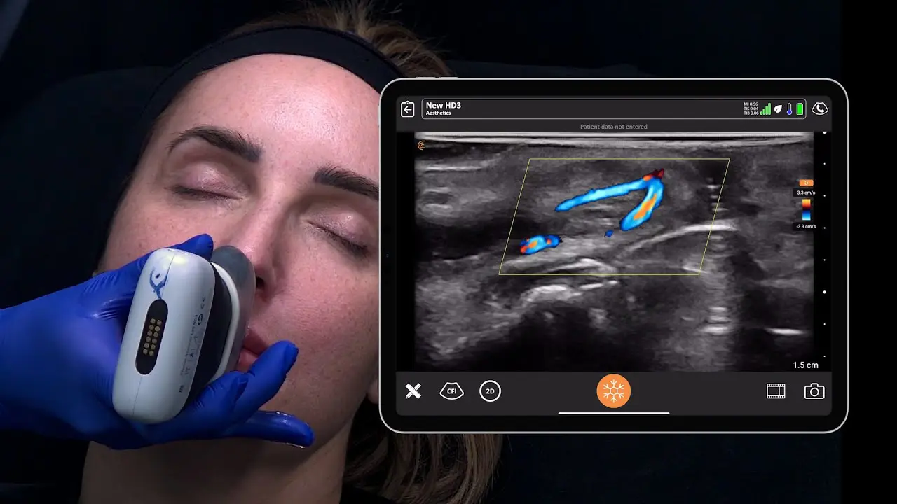
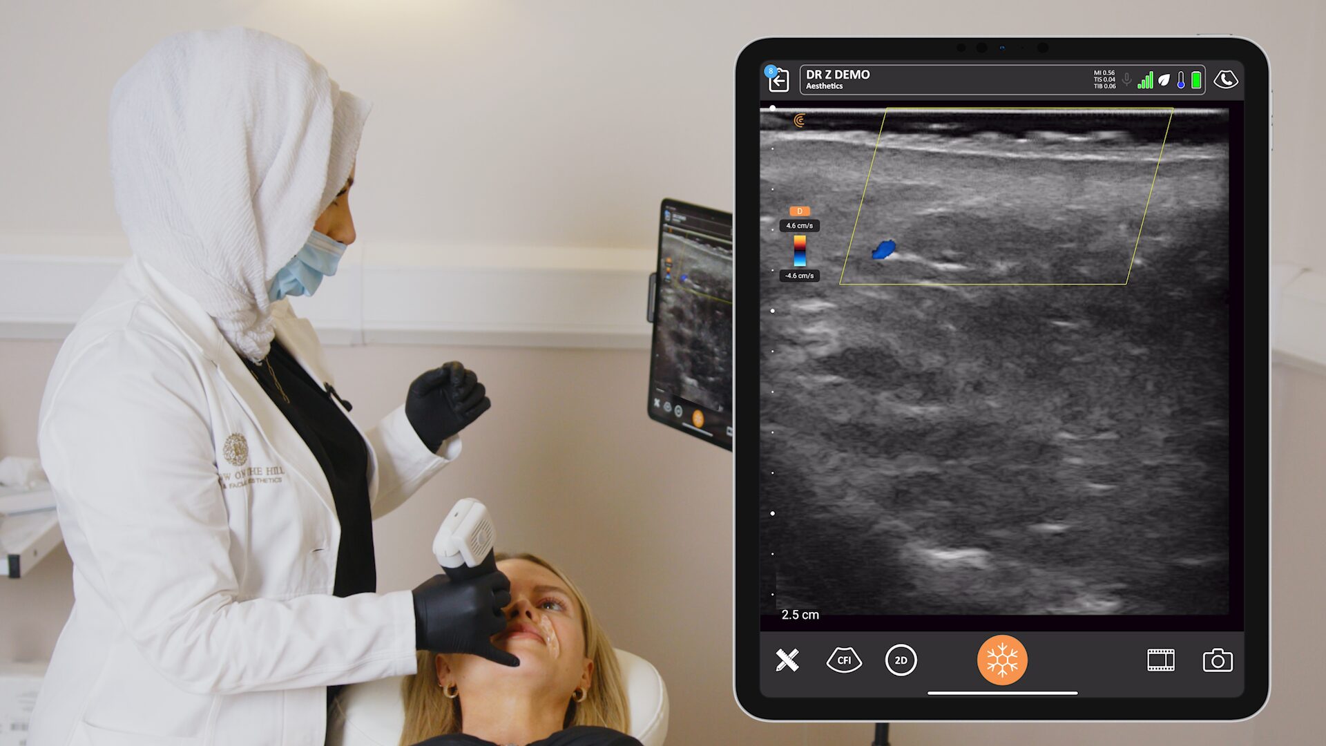
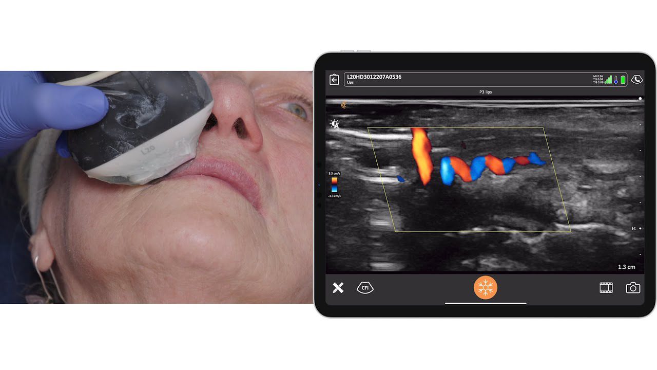
Vascular Safety: Dr. Ravera emphasizes that anatomical variations in vessels and foramina are the rule, not the exception. «I want to highlight that this area is one of the most dangerous zones in the face, because our patients can become blind. And these arteries or bundles are the most variable in our patients». Therefore, she adds: «the only way to account for variability is to use imaging, in this case, ultrasound examination». The foramina appear as an interruption in the hyperechoic line of the bone’s cortex, and a neurovascular bundle will always be found at this site.
SLEB as a Biomarker: The subepidermal low-amplitude band (SLEB) is a crucial ultrasound biomarker that indicates a functional and structural alteration of the papillary dermis. It appears as a hypoechoic band beneath the epidermis.
«SLEB is a very important ultrasonography biomarker. It is objective, valid and reproducible», Dr. Ravera note. “It is useful for assessing skin aging and conditions such as atopic dermatitis, psoriasis, and cutaneous lymphomas.”
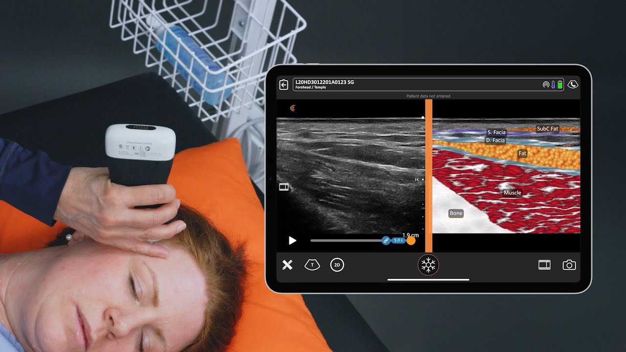
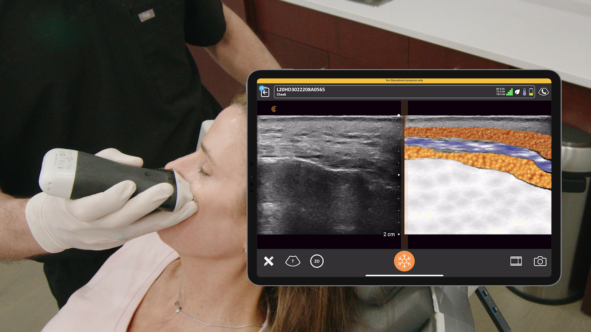
Understanding Fat Compartments: Ultrasound allows clinicians to differentiate between superficial and deep fat compartments, which appear with different echogenicity and mobility on the scan. Dr. Ravera points out that Clarius T-Mode feature is particularly helpful for new users – “it colors and texturize very well the different structures of the temple».
In this 2-minute video, Dr. Ravera demonstrates the ultrasound appearance and thickness of the SLEB. The SLEB, or subepidermal low-echogenic band, is thought to be a marker of skin aging.
Specialized Ultrasound for Safer Procedures and Better Results
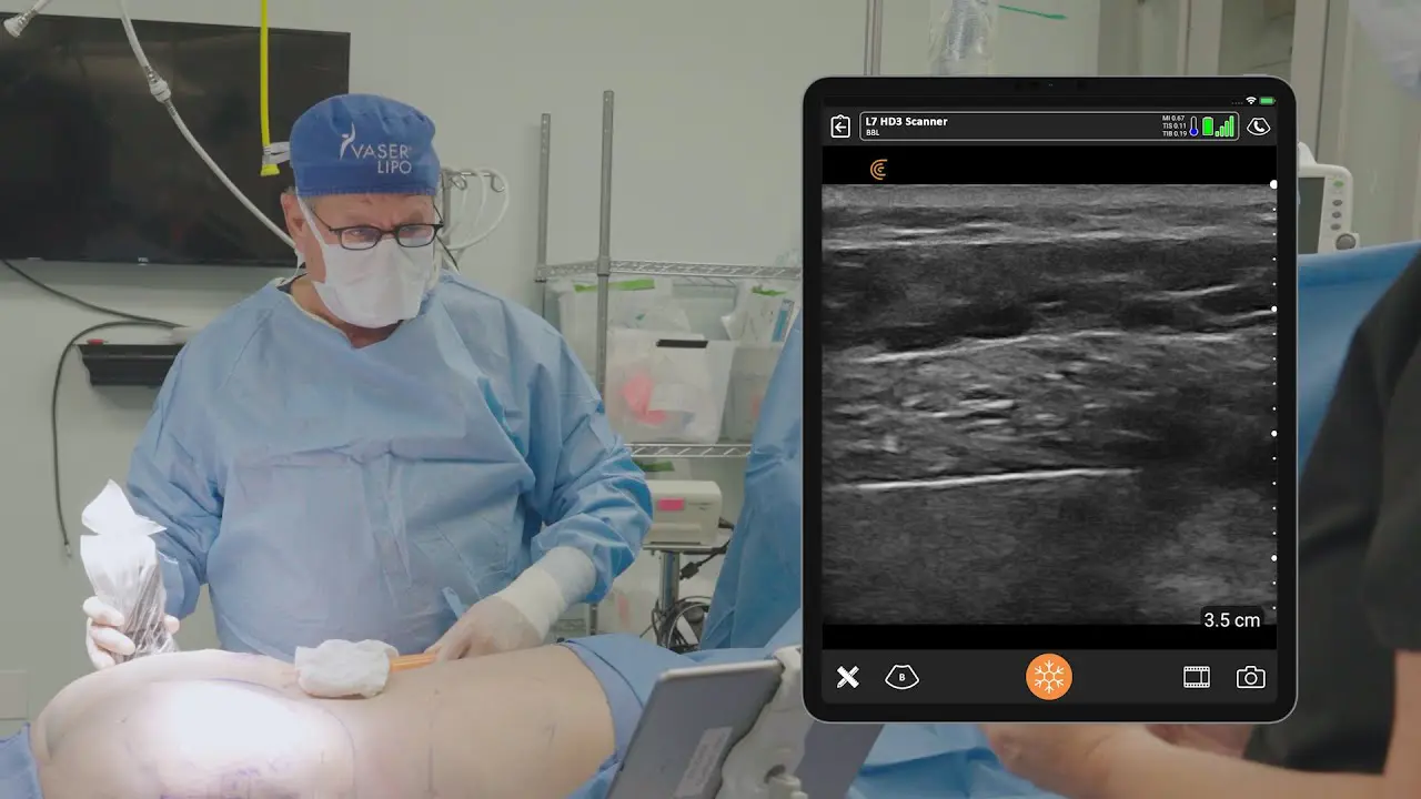
Clarius ultrasound has enabled more aesthetic clinicians to use ultrasound guidance by making high-frequency imaging easy and affordable, with image quality that rivals traditional systems but at a fraction of the cost. The wireless, handheld units are ideal for superficial imaging, providing high-definition views of skin, muscles, vessels, fascia, and foramina.
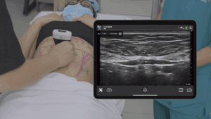
Clarius T-Mode is a standout feature. It’s an innovative AI-powered tool that colorizes and texturizes different tissue layers, allowing for quick and easy differentiation. «It really does help,» said Janaye Smith, a sonographer who demonstrated T-Mode in a live scanning session during the webinar. This is particularly useful for distinguishing between muscle and fat, which can look similar in a B-mode scan.
Watch this 5-minute live demonstration from the Webinar to see T-Mode in action.
Watch the full webinar to learn more about facial ultrasound anatomy! Visit Clarius Classroom for more short tutorials featuring Dr. Ravera and other aesthetic experts.
Improve Patient Safety and Procedure Outcomes with Ultra-High-Definition Ultrasound for Facial Aesthetics
Clarius handheld ultrasound is the leading choice for aesthetics practitioners like Dr. Ravera to clearly visualize facial and superficial anatomy in real-time to safely guide procedures. With exceptional superficial imaging, the new advanced aesthetic protocol, and new T-Mode, the Clarius L20 HD3 is the popular choice for facial aesthetics.
Learn more about Clarius AI-powered ultrasound on the aesthetics specialty page. Or contact us for a personalized virtual demonstration.
