


For primary care providers, the gap between a physical exam and a definitive diagnosis often means a referral and a wait—a gap that point-of-care ultrasound (POCUS) is now closing right at the bedside. During a recent webinar, Dr. Tatiana Havryliuk—Emergency Physician and founder of Hello Sono—shared her roadmap for integrating ultrasound into everyday practice.
Dr. Havryliuk knows the versatility of these devices firsthand. As she noted during the presentation, while showing a photo of herself scanning a patient on a glacier:
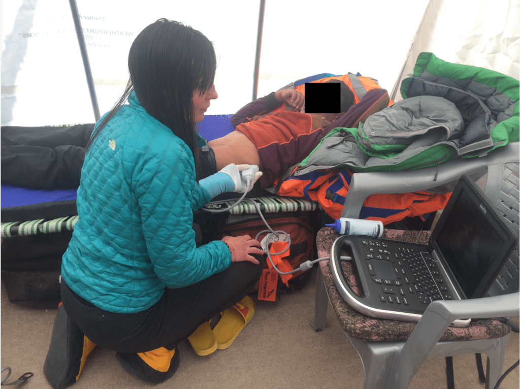
This is actually me at 17,000ft at Everest Base Camp in Nepal performing point of care ultrasound… there’s no reason why we can’t make this happen for your primary care practice.»
Watch the full webinar to see Dr. Havryliuk’s deep dive into scan techniques and live demonstrations. Read on for some highlights and expert tutorials.
Why POCUS? Why Now?
Adoption of POCUS in primary care is growing, particularly in rural areas where radiology support is scarce. Dr. Havryliuk highlights why now is the perfect time to start:
- Affordability: «For about $5,000, you can get… a handheld device that lets you do all the basic applications at the bedside… In the past, you had to spend $40,000 or more to have a cart-based system.»
- Patient Trust: «95% of patients who were scanned during a clinic visit actually thought that their level of service was better, and 65% of them reported that they trusted their provider more.»
- Efficiency: You get an extra data point immediately, allowing you to start treatments—like diuresis or antibiotics—sooner, rather than waiting for X-ray results.
5 High-Yield POCUS Applications for Primary Care
Dr. Havryliuk detailed five core exams that are high-yield and easier to learn than advanced applications.
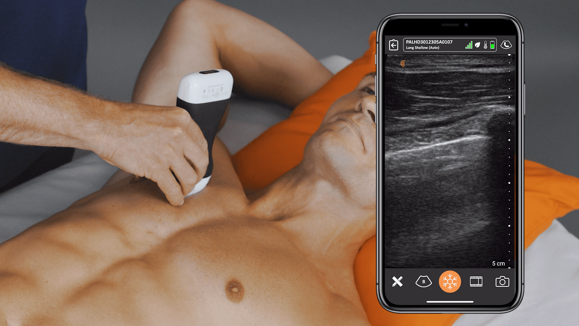
1. Lung Ultrasound
This exam helps rule out dangerous pathology immediately for patients with shortness of breath, chest pain, or cough.
- Pneumonia vs. Atelectasis: Dr. Havryliuk explained the frustration of ambiguous radiology reports: «When you get that chest x ray read that says ‘clinically correlate atelectasis versus pneumonia’… [with] ultrasound you can actually tell, ‘Hey, this is pneumonia because I’m seeing those dynamic air bronchograms.'»
- Pneumothorax: Look for «lung point,» which Dr. Havryliuk notes is «100% specific for a pneumothorax.»
VIDEO: Lung Ultrasound Techniques for Primary Care
In this video, Dr. Havryliuk describes indications for lung ultrasound and the benefits of lung ultrasound in a primary care setting. She uses the Clarius PAL HD3 for the exam.
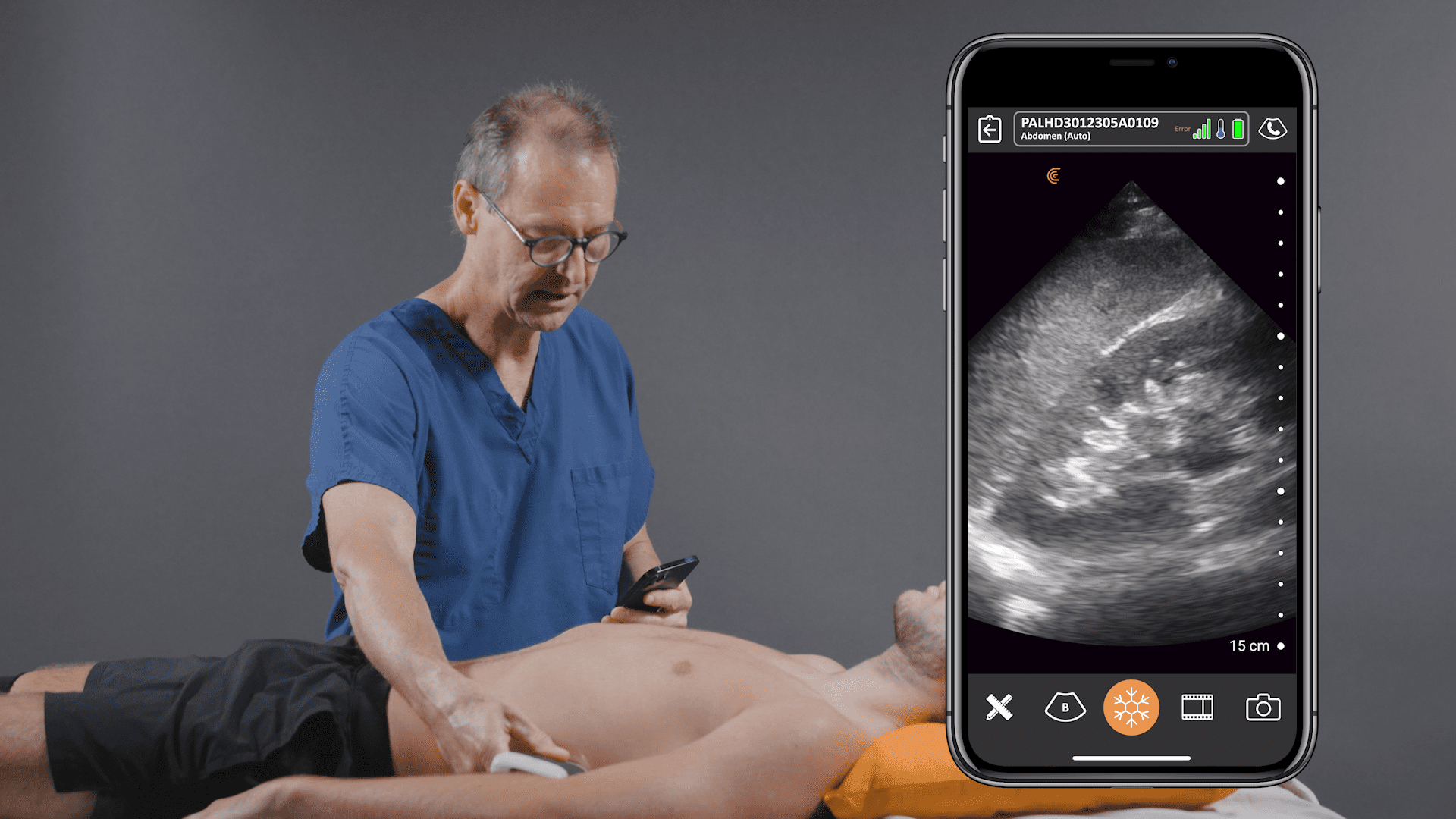
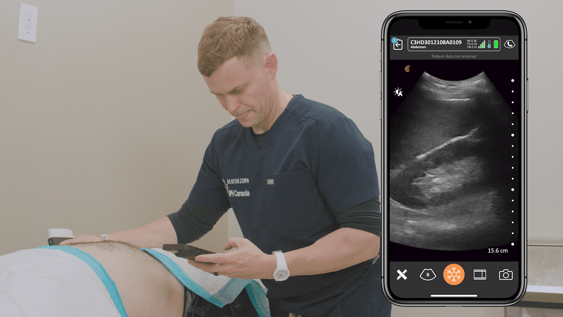
2. Abdominal Aorta (AAA) Screening
Essential for screening men over 65 who smoke, or evaluating abdominal pain.
- Technique: Visualize the aorta in transverse and sagittal planes.
- Crucial Tip: «Make sure you are measuring outer to outer wall of the aorta so you don’t miss an aneurysm.»
- Dissection Warning: «Just remember, do not use POCUS to rule out a dissection. You can only rule it in with POCUS.»
VIDEO: POCUS of the Abdominal Aorta
Watch Dr. Havryliuk demonstrate how to scan the abdominal aorta to rule out AAA in patients presenting with abdominal, flank, or low back pain. She uses the Clarius PAL HD3 for the exam.
3. Renal and Bladder
Ideal for workups of hematuria, flank pain, or urinary retention.
- Hydronephrosis: Look for anechoic (black) fluid in the center of the kidney.
- Stones: Ultrasound can identify stones before they move; Dr. Havryliuk pointed out hyperechoic specks in a scan as stones «that have not traveled yet… about to wreak havoc.»
VIDEO: Renal and Bladder Ultrasound
In this video, Dr. Havryliuk demonstrates her ultrasound techniques for a quick ultrasound assessment to rule out obstruction and urinary retention in patients presenting with suprapubic pain, difficulty voiding, or flank pain.
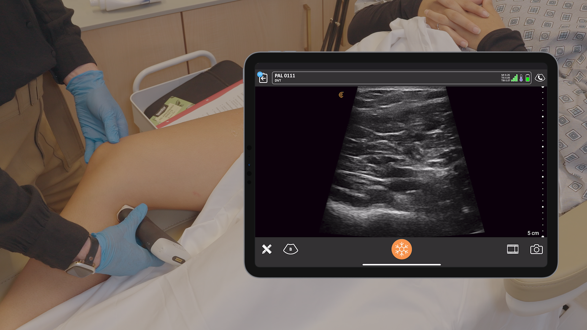
4. Lower Extremity DVT
POCUS allows you to rule out proximal Deep Vein Thrombosis (DVT) at the bedside.
- The «Pop on Top» Mnemonic: When scanning the popliteal vein, remember it «sits on top of the artery… pop on top if you want to remember it pretty easily.»
- Patient Reassurance: «Instead of having patients, ‘Hey, hey, wait for three days or go to the E.R.’… you get to tell them at the bedside that you’re not seeing a proximal leg DVT.»
VIDEO: Lower Extremity DVT Ultrasound
Ultrasound is the standard for diagnosing DVT and can help prevent a potentially dangerous pulmonary embolism if a clot is found. In this video, Dr. Havryliuk demonstrates her 3-point compression technique to assess the deep veins of the legs.
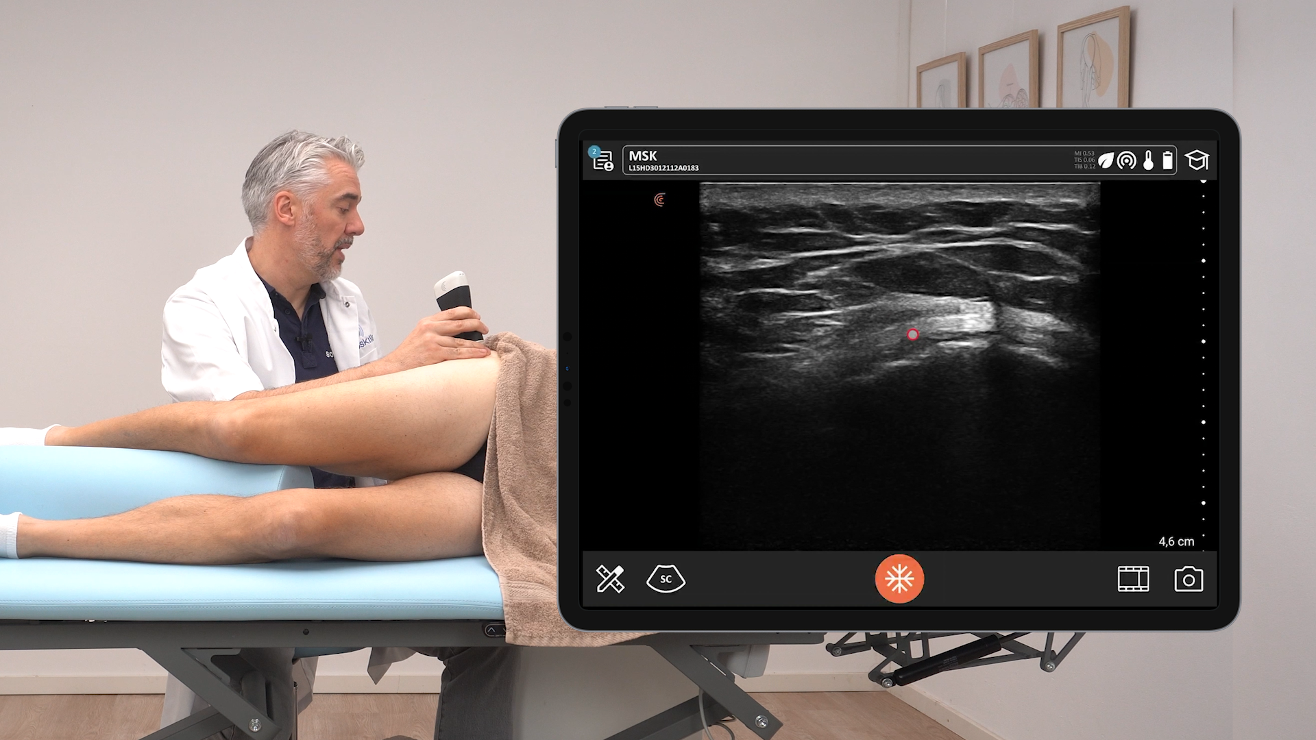
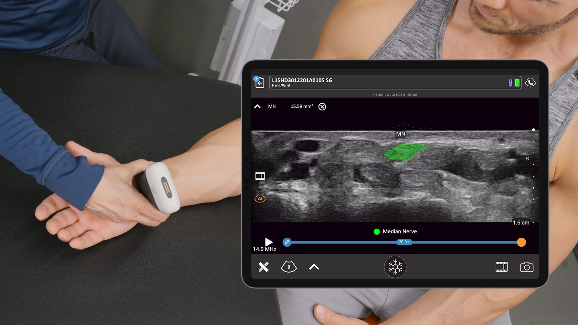
5. Musculoskeletal (MSK)
Useful for swollen joints or tendon injuries.
- Safety: You can avoid radiation from unnecessary X-rays.
- Procedure Guidance: It ensures you aren’t «doing knee taps on patients who have no fluid in their knee.»
VIDEO: Achilles Assessment
Watch this video to see Dr. Havryliuk demonstrate how to perform a POCUS assessment of the Achilles tendon in patients presenting with posterior ankle pain.
Implementation: Building a Compliant Program
To successfully implement POCUS, Dr. Havryliuk emphasizes that you cannot just buy a scanner and start; you need a workflow.
- Credentialing is Key: «I do believe that credentialing is necessary… and it does take some time. I would estimate 3 to 12 months at the very least.»
- Proficiency Standards: She recommends a minimum of 25 supervised scans per application to achieve proficiency.
The ROI of POCUS – for clinics in the United States
Dr. Havryliuk broke down the financial benefits for a primary care clinic in the United States that could result from direct billing and indirect savings.
- Revenue: A primary care clinic performing 10 exams per week can generate between $30,000 and $90,000 per year in billable revenue.
- Value-Based Care: POCUS fits perfectly into value-based models by delivering patient-centric, cost-effective treatment.
Q&A with Dr. Tatiana Havryliuk
Q: Do you use Doppler flow while doing a DVT study?
Dr. Havryliuk: «Typically for the compression-focused study, you don’t need to use Doppler… I don’t usually use it unless I am having a hard time visualizing the vessels… compression is the most important part of this exam.»
Q: How do you differentiate between acute and chronic DVT?
Dr. Havryliuk: «With an acute clot… the clot will look more anechoic than hyper echoic. And it’s also not going to be like attached to the wall… versus on the chronic ones, you might have a partial clot that’s even a little bit calcified. Sometimes they are like attached to the wall of the vessel.»
Q: Is it risky to do a DVT scan if you aren’t certified?
Dr. Havryliuk: «I would get your scans… get CME, get your scans reviewed, have documentation. I think that would be a huge protection medical legally.»
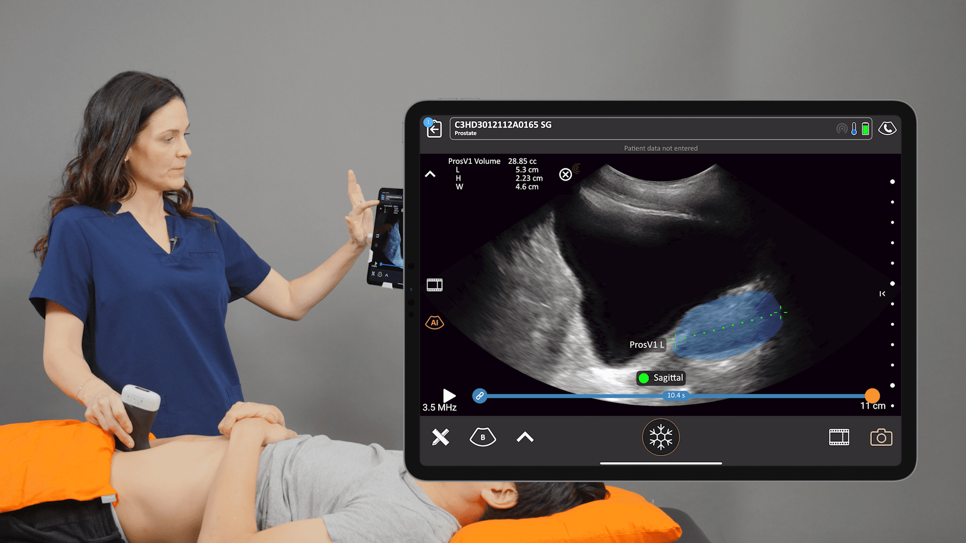
Why Clarius is the Ideal Choice for Primary Care
During the live demonstration, Patrick Villaruel, Clarius Clinical Specialist, highlighted why the Clarius HD3 is specifically suited for the primary care environment:
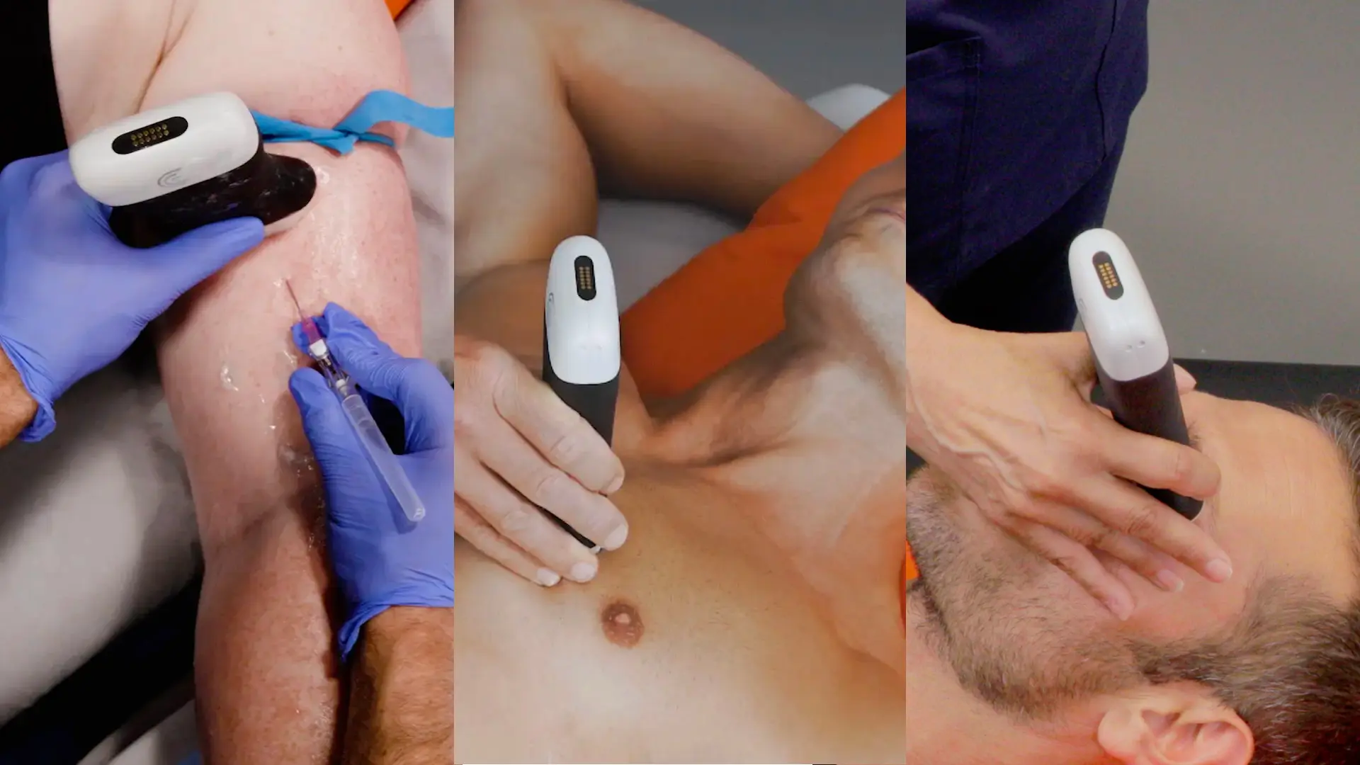
- Versatility (The PAL Probe): Patrick demonstrated the «Phased Array Linear» (PAL) probe, which combines two probes in one, allowing you to scan deep structures (like the bladder) and superficial structures (like the shoulder) without changing devices.
- AI-Powered Assistance: Features like Voice Controls allow hands-free adjustments, while AI Bladder automatically measures volume, increasing exam efficiency.
- Educational Overlay: The «T-Mode™ acts like a live biology textbook, labeling tissues in real-time to help learners identify anatomy.
- Seamless Workflow: Clarius is wireless, works with iOS and Android, and includes Clarius Cloud for unlimited exam storage and reporting from anywhere.
Ready to start your POCUS journey?
Watch the full webinar to see live scanning demonstrations and to get detailed explanations of how Dr. Havryliuk uses ultrasound to deliver the best patient care. When you’re ready to consider Clarius for your practice, contact us for a personal virtual demo to learn more.


