In the world of feline medicine, point-of-care ultrasound (POCUS) is rapidly gaining recognition as an invaluable diagnostic tool. POCUS offers several advantages, including rapid diagnosis, non-invasive visualization, and serial monitoring capabilities.
To delve into this topic for our veterinary webinar series, we invited back Dr. Soren Boysen, Professor, Faculty of Veterinary Medicine at University of Calgary, and Dr. Serge Chalhoub Associate Professor, Faculty of Veterinary Medicine, University of Calgary. Hundreds of veterinarians tuned in to the webinar: Veterinary POCUS: Diagnosing Renal Pelvic Dilation, to learn proven ultrasound techniques from the popular dynamic duo.
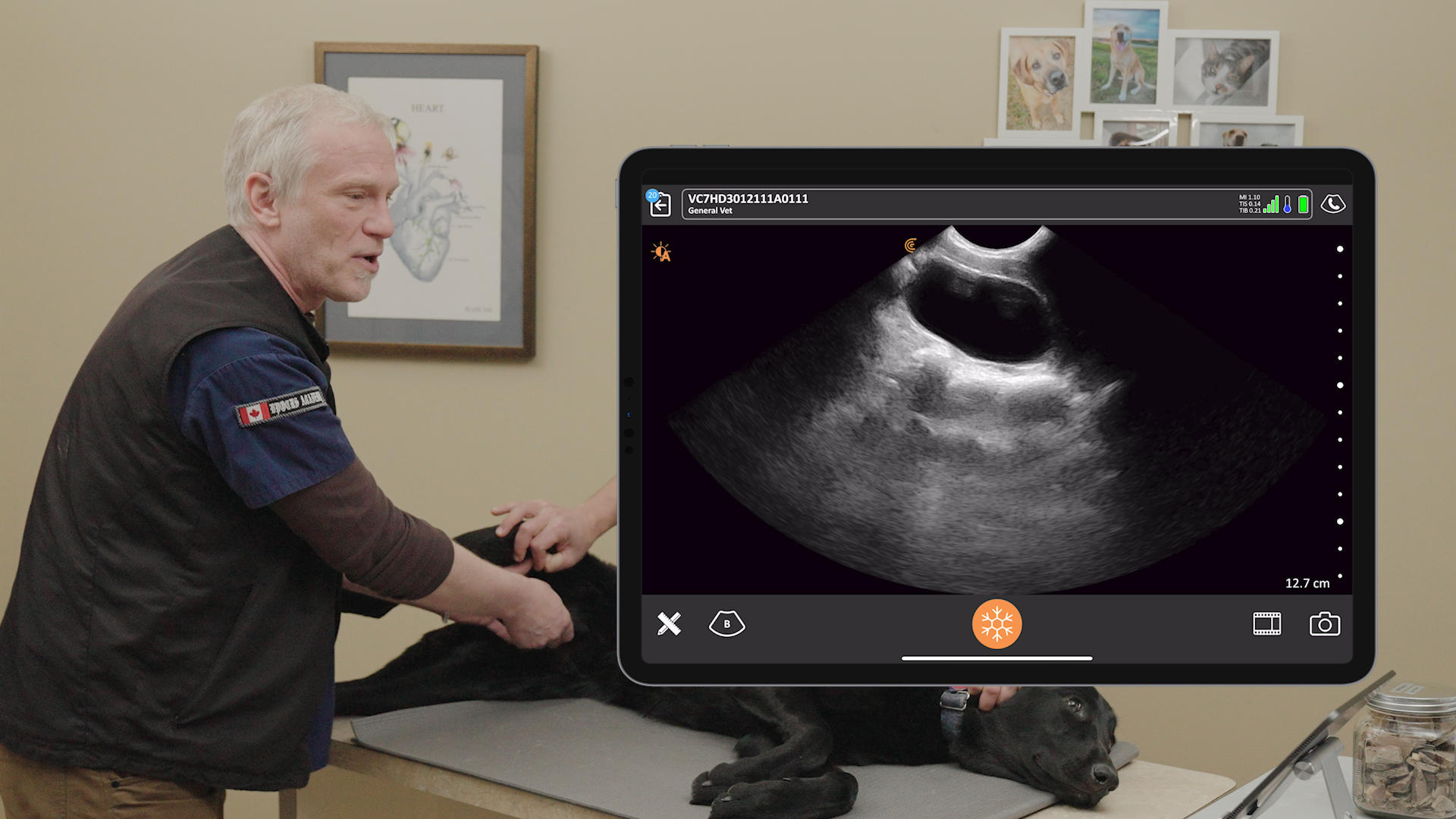
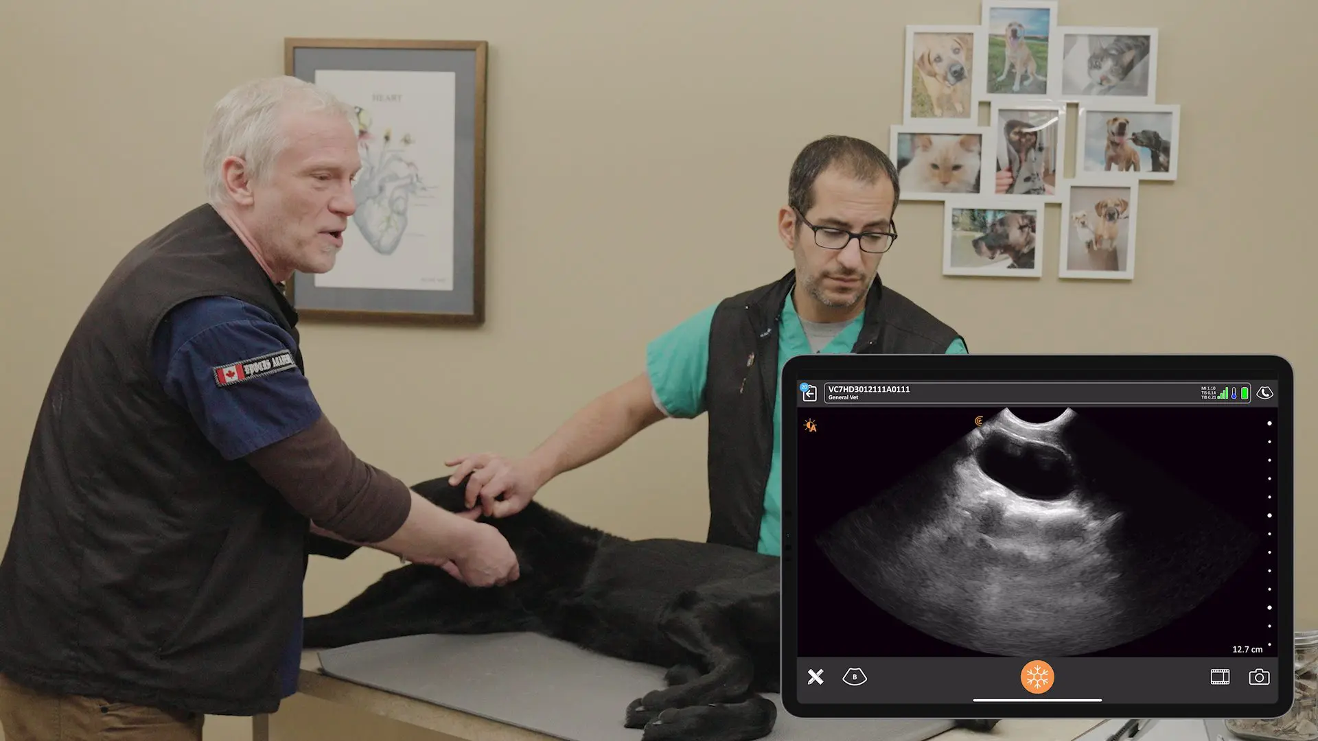
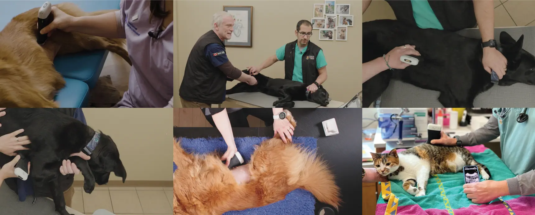
During the one-hour webinar, Dr. Boysen and Chalhoub use feline exams captured on video with Daisy, a large domestic short-haired cat to explain the role of POCUS in identifying renal pelvic dilation in cats; they demonstrate ultrasound techniques to evaluate the kidneys at the right and left paralumbar sites, and they teach how to narrow the differential diagnosis for renal pelvic dilation in cats.
Watch the webinar at your convenience. It’s RACE-approved for 1 CE/CPD Credit if you access it through the Vet Show. Or simply scroll for key highlights.
Key Applications of POCUS in Feline Azotemia:
One of the key applications of POCUS in cats is the assessment of acute azotemia, a condition characterized by elevated levels of urea and creatinine in the blood. POCUS can help differentiate between pre-renal, renal, and post-renal causes of azotemia, allowing for prompt and targeted treatment.
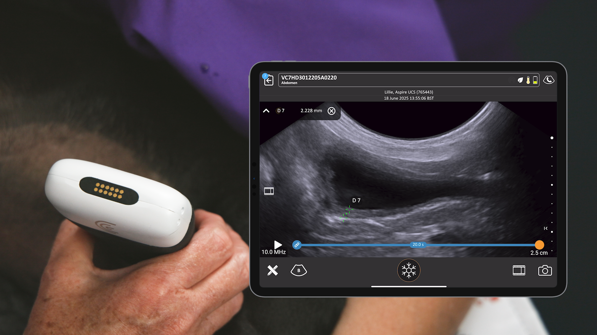
- Assessment of renal pelvic dilation: POCUS can accurately measure the degree of renal pelvic dilation, aiding in the diagnosis of ureteral obstructions. A dilated renal pelvis greater than 13 millimeters in diameter is highly suggestive of ureteral obstruction in cats.
- Evaluation of urine production: Serial POCUS examinations of the urinary bladder can help track urine volume production, providing valuable information about the cat’s response to treatment.
- Assessment of volume status: POCUS can be used to evaluate the cat’s hydration status and guide fluid therapy decisions. By visualizing the caudal vena cava and heart, veterinarians can determine if a fluid bolus is necessary and monitor for signs of fluid overload.
Tips for Successful Feline POCUS:
- Patient positioning: Scan the cat in a position that is most comfortable for them, such as lateral recumbency or sternal recumbency.
- Probe placement: Use anatomical landmarks, such as the subxiphoid region and the paralumbar sites, to guide probe placement.
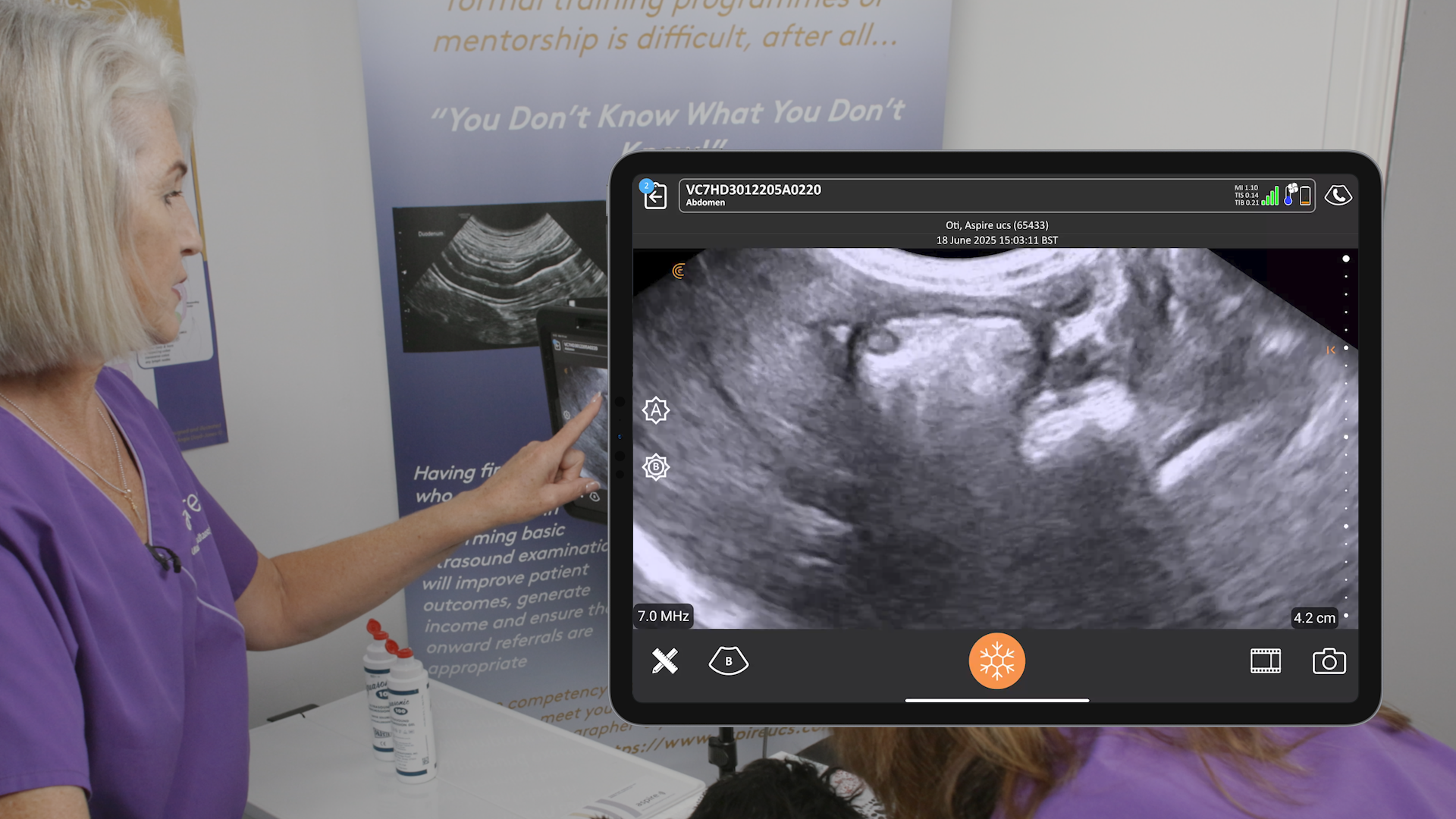
- Fanning technique: Fan the probe through all planes in both long and short axis to ensure thorough visualization of the kidneys and surrounding structures.
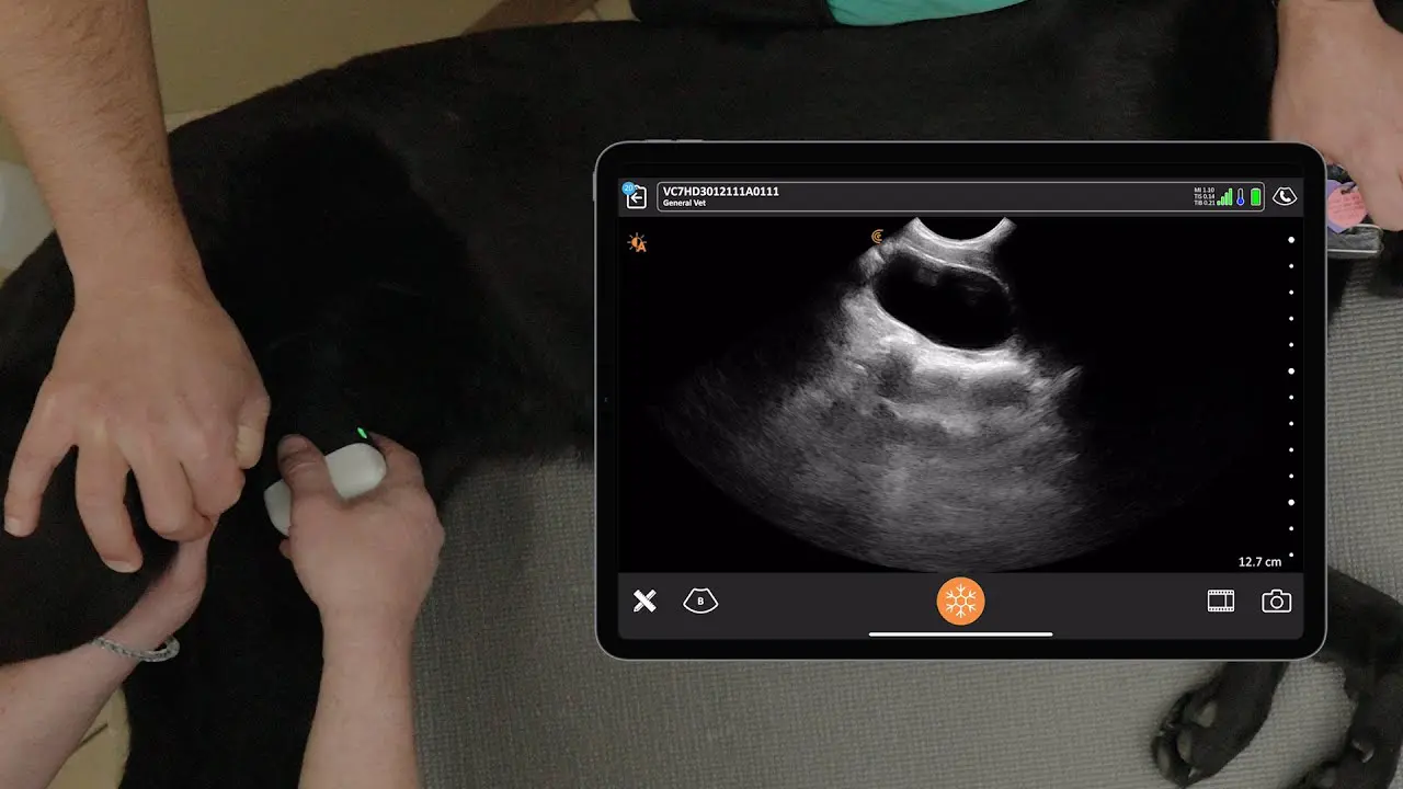
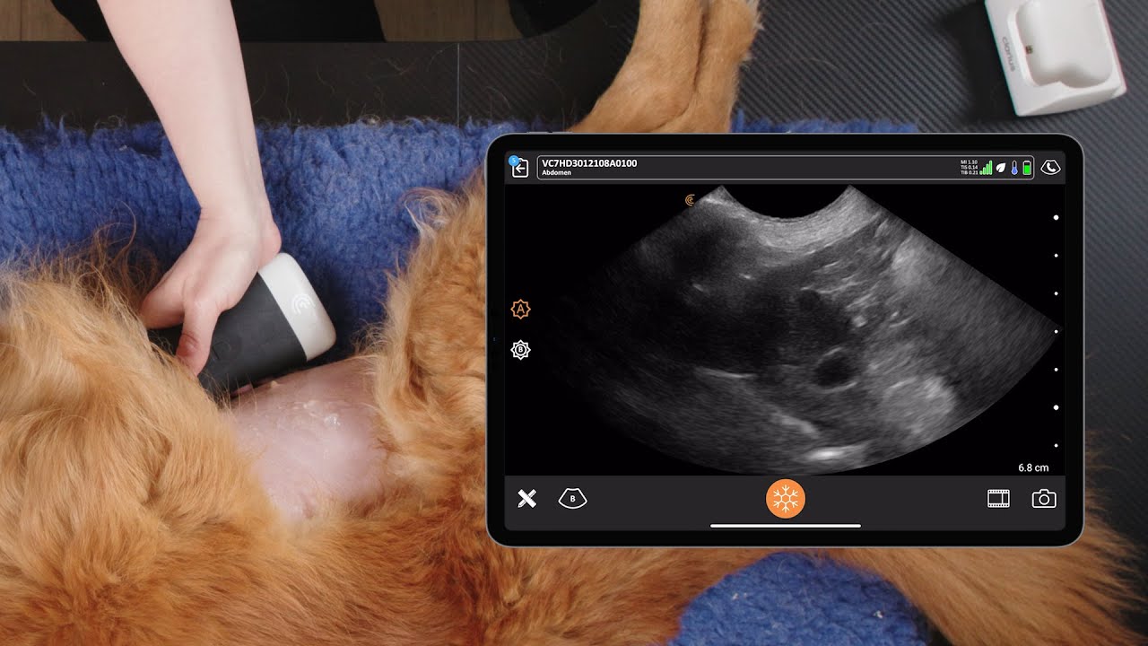
VIDEO: Scanning the Left Kidney Paralumbar Site with Handheld Ultrasound
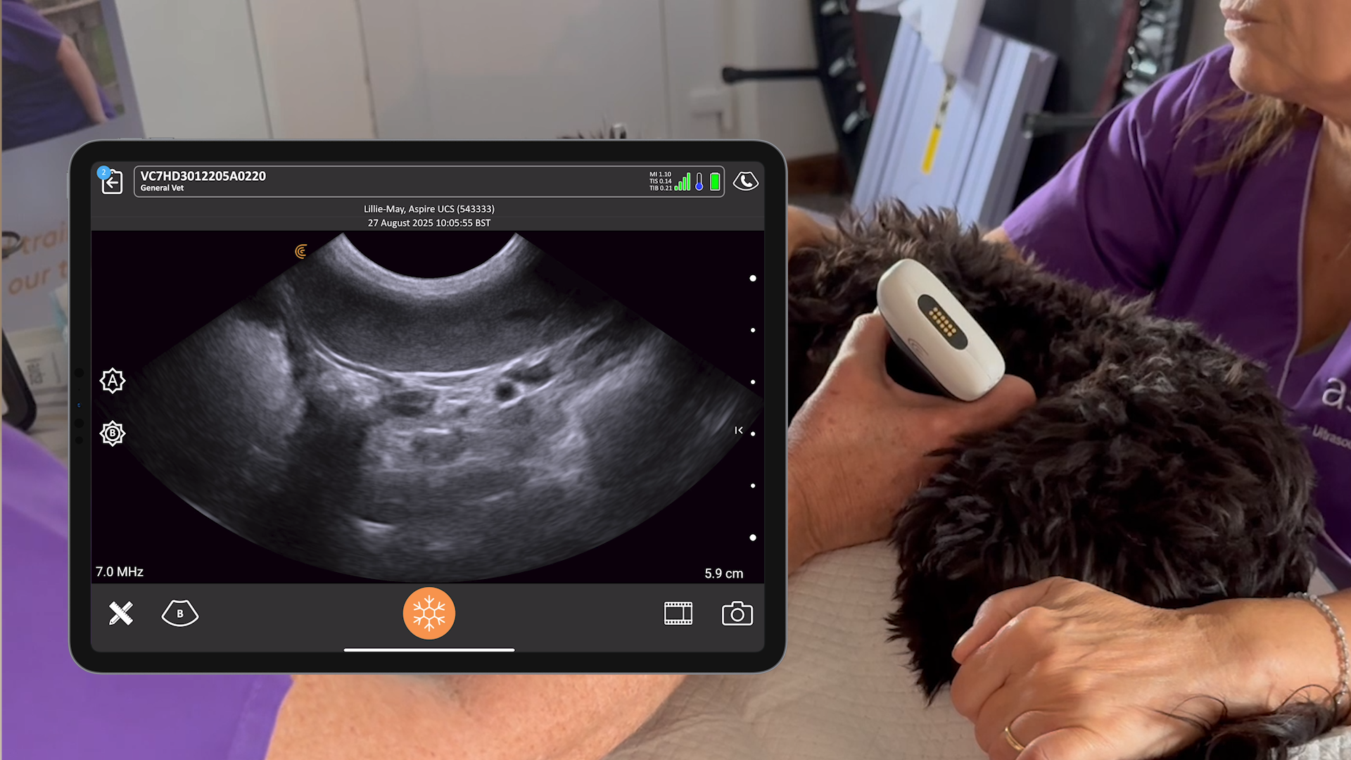
Watch Dr. Chalhoub in action in this 3-minute video as he uses the Clarius C7 Vet HD3 to locate the left kidney in the left paralumbar space to rule out renal pelvic dilation.
To Scan or Not to Scan for a Triage Exam
During a triage exam, we rarely shave. If you part the fur so that you can put alcohol right on the skin, that’s going to be pretty darn good contact. It’s a focal window and therefore we get away with it. If you think about it, the goal of clipping is to remove the fur to prevent air from getting trapped. If you separate the fur and can see the skin, you’re going to get a similar effect. It would be different for a full abdominal ultrasound exam.
Find answers under the fur with high-definition wireless ultrasound
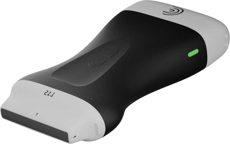
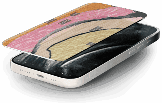
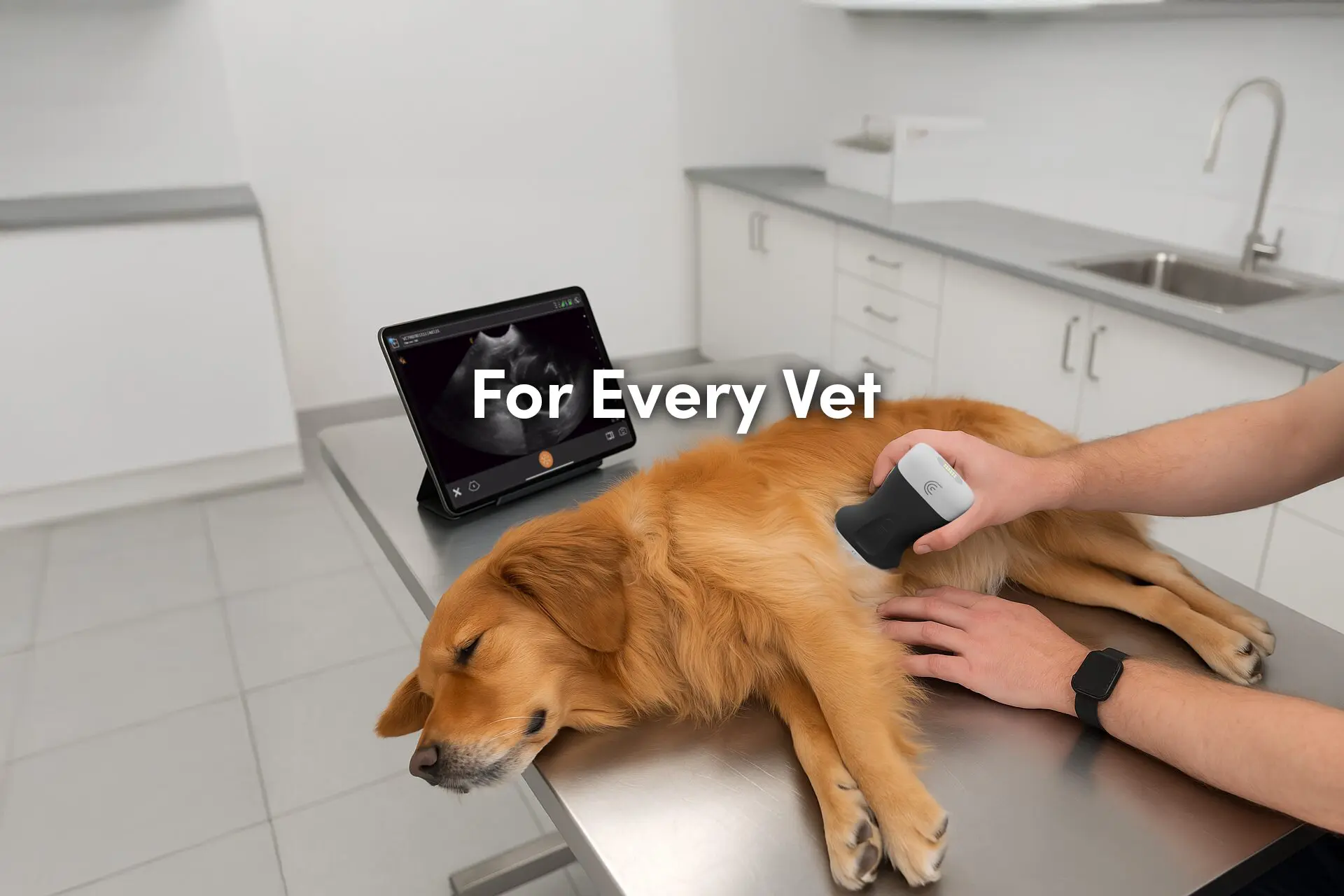
POCUS is a powerful tool that can significantly enhance the diagnosis and management of feline kidney problems. Now you can optimize comfort with on-the-spot imaging and no wires to startle your furry patients. Ultra-portable and affordable, Clarius Vet HD3 delivers the quality imaging and performance of traditional systems without complex knobs and buttons. The AI-powered app is as easy to use as your smartphone.
We invite you to book a virtual demonstration to discuss which Clarius scanner is ideal for your veterinary practice. Or visit our Veterinary Specialty page to learn why Clarius ultrasound is a popular choice for veterinarians shopping for an ultrasound system.
