



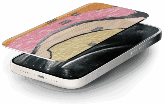
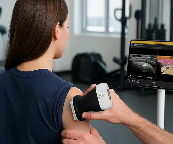
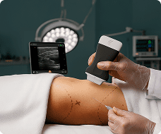
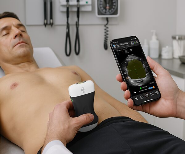


New to lung ultrasound? Clarius T-Mode Lung uses AI-driven anatomical overlays to turn everyday POCUS scans into an interactive learning tool. By displaying a live B-mode image side-by-side with a color-coded tissue overlay, T-Mode helps clinicians and students quickly identify key thoracic structures and build confidence.
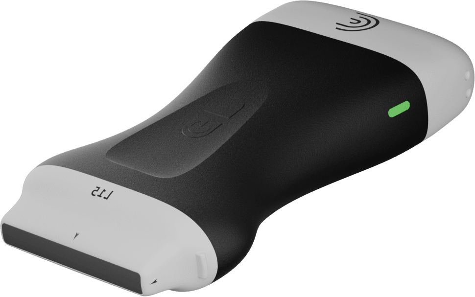
Compatible Scanners: Clarius C3, PAL, L7, and L15 scanners (using the Lung Preset).
Key Features & Workflow:
Sagittal Placement: Position the scanner in the long-axis plane to visualize tissue layers from the dermis to the pleural space.
Side-by-Side Visual Overlay: Clearly differentiate between dermis, subcutaneous tissue, muscle, ribs (with diagnostic acoustic shadows), and lung tissue.
Interactive Live Labels: Toggle anatomical labels on or off to quiz students or test your own knowledge.
Opacity Adjustment: Slide to adjust grayscale opacity for optimum image contrast and anatomical clarity.
Cloud Integration: Freeze, capture, and automatically save both B-mode and labeled T-Mode scans directly to the Clarius Cloud.
