


In the evolving landscape of musculoskeletal (MSK) care, physiotherapists are continually seeking innovative tools to refine their diagnostic capabilities and optimize patient outcomes. Musculoskeletal ultrasound (MSK US) stands out as a powerful, non-invasive imaging modality that offers real-time, dynamic insights into soft tissue and bony structures, revolutionizing how physiotherapists assess and manage a wide range of conditions.
As highlighted by renowned educator Marc Schmitz, founder and CEO of Sonoskills, during a recent Clarius webinar, “Ultrasound is a fantastic tool to look at this wrist and this hand region.” This sentiment extends across the entire musculoskeletal system, offering physiotherapists an unparalleled view into the body’s intricate mechanics.
Dynamic Visualization in Clinic: Seeing Beyond Static Images
One of the most significant advantages of MSK US is its ability to provide dynamic, real-time imaging. Unlike static imaging modalities such as X-rays or MRI, ultrasound allows clinicians to observe structures in motion, during stress tests, and throughout the range of movement. Marc emphasizes this, stating, “This functional real-time scanning, assisting in your diagnosis, and your clinical reasoning is truly magnificent.” This dynamic capability is crucial for identifying subtle instabilities, impingements, or abnormal gliding patterns that might be missed with static imaging.
For instance, in cases of intersection syndrome in the wrist, ultrasound enables the visualization of friction and irritation between tendon compartments during movement. Marc notes, “The main value of the ultrasound here is that you can do functional scanning so you can really see how these two layers interact with each other during movement.” This allows for precise identification of the problem’s source and helps in tailoring targeted interventions.
Watch the free one-hour webinar with Marc Schmitz at your convenience: POCUS for MSK: Mastering Hand and Wrist Ultrasound. Read on for quick highlights and video demonstrations of hand and wrist ultrasound with the Clarius L15 HD3.
Elevating Care: Tangible Benefits of MSK US for Physiotherapists
Integrating MSK ultrasound into physiotherapy practice offers a multitude of benefits, directly impacting diagnostic accuracy, treatment planning, and patient engagement:
- Enhanced Diagnostic Accuracy: Ultrasound provides high-resolution imaging of soft tissues, including tendons, ligaments, muscles, and nerves, allowing for precise identification of pathologies like tears, inflammation, and effusions. As Marc explains, “Ultrasound does well at depicting the system, the soft tissue, and articular structures.”
- Real-Time Functional Assessment: The ability to visualize structures in motion is invaluable for assessing dynamic pathologies, such as tendon snapping, joint instability, or impingement syndromes. “You can also provoke certain movements to see in real time what’s happening during stressful movements,” says Marc.
- Immediate Feedback and Patient Education: Patients can observe their own anatomy and pathology in real-time, fostering a deeper understanding of their condition and promoting active participation in their rehabilitation. This visual feedback can significantly improve patient compliance and satisfaction.
- Guidance for Interventions: Ultrasound can guide various interventions, from dry needling to injections, ensuring precise targeting of affected tissues and minimizing risks. “Fluid is your friend,” Marc states, referring to fluid collections that can be targeted during interventions.
- Monitoring Treatment Progress: Regular ultrasound scans can objectively track changes in tissue healing, reduction in inflammation, or improvements in tendon gliding, allowing physiotherapists to adapt treatment plans as needed.
- Early Detection of Fractures: While X-rays are often the first line for suspected fractures, ultrasound can play a crucial role in early detection, especially for conditions like scaphoid fractures that may be initially missed on X-ray. Marc highlights, “X-ray imaging is not always suitable for scaffold fractures, and ultrasound can really help in this early stage.”
- Differentiation of Tissue Types: Ultrasound’s ability to distinguish between different tissue densities (from anechoic fluid to hyperechoic bone) aids in accurately characterizing pathologies. “Every anatomical tissue has its specific gray value,” Marc explains.
Case Examples of Diagnosing Hand and Wrist Pathologies with Ultrasound
The webinar showcased several compelling examples where ultrasound proves invaluable:
Intersection Syndrome: Differentiating between proximal and distal intersection syndromes by observing the friction and swelling of specific tendon compartments (e.g., extensor compartment 1 over 2, or EPL over ECRB/L).
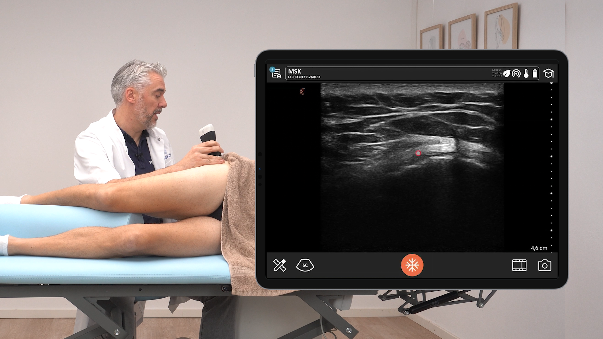
Watch this video to see Marc demonstrate his technique for imaging the extensor tendons in compartment 2 proximal and distal to Lister’s Tubercle.
Flexor Tendon Pulley Injuries: Assessing pulley thickening and tears, which can lead to “trigger finger” or “bowstringing” of tendons. Marc notes that for trigger finger, “ultrasound is the preferred imaging technique, and especially due to the possibility to do functional scanning.” The measurement of tendon-to-bone distance is critical here.
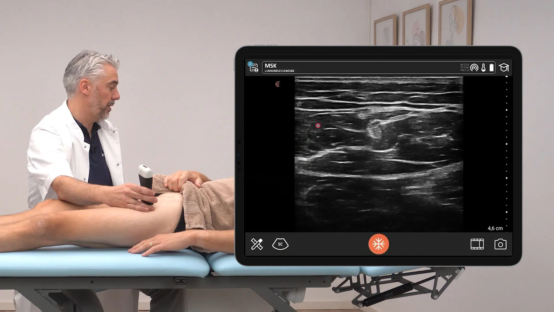
Flexor pulley injuries are often seen in rock climbers, and high-resolution ultrasound can be used to assess the pulleys. Watch this video demonstration of the flexor pulleys and surrounding structures.
Scaphoid Fractures: Identifying cortical irregularities and step-off deformities in the scaphoid bone, particularly in cases where X-rays are inconclusive. Marc emphasizes that ultrasound is “more sensitive and specific than X-ray imaging for ASC for fractures.”
Ultrasound of the scaphoid bone can detect small fractures that can be missed on X-ray. Marc demonstrates how to assess the scaphoid with both dorsal and palmar approaches in this video.
Scapholunate (SL) Ligament Instability: Visualizing gapping in the scapholunate joint during dynamic stress tests, indicating a tear or instability.
In this video, Marc Schmitz demonstrates how to assess the scapholunate ligaments, a commonly injured ligament in the wrist.
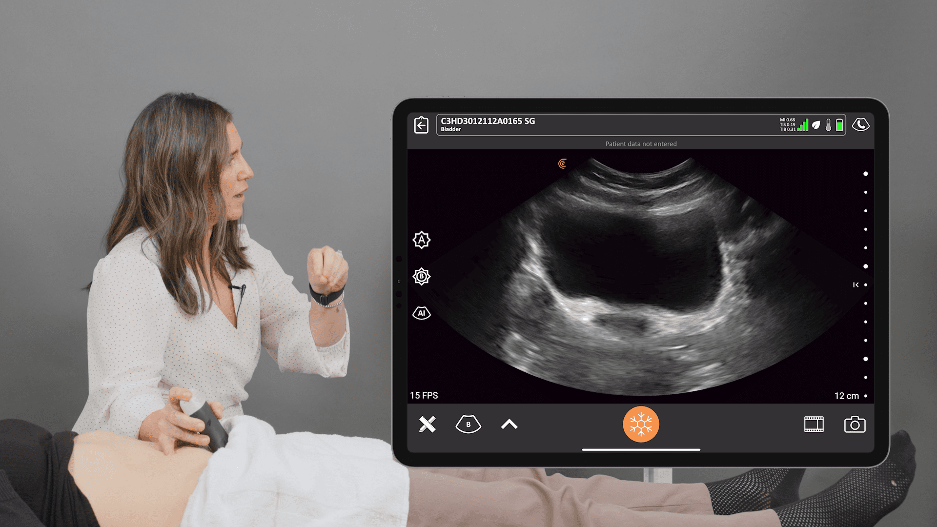
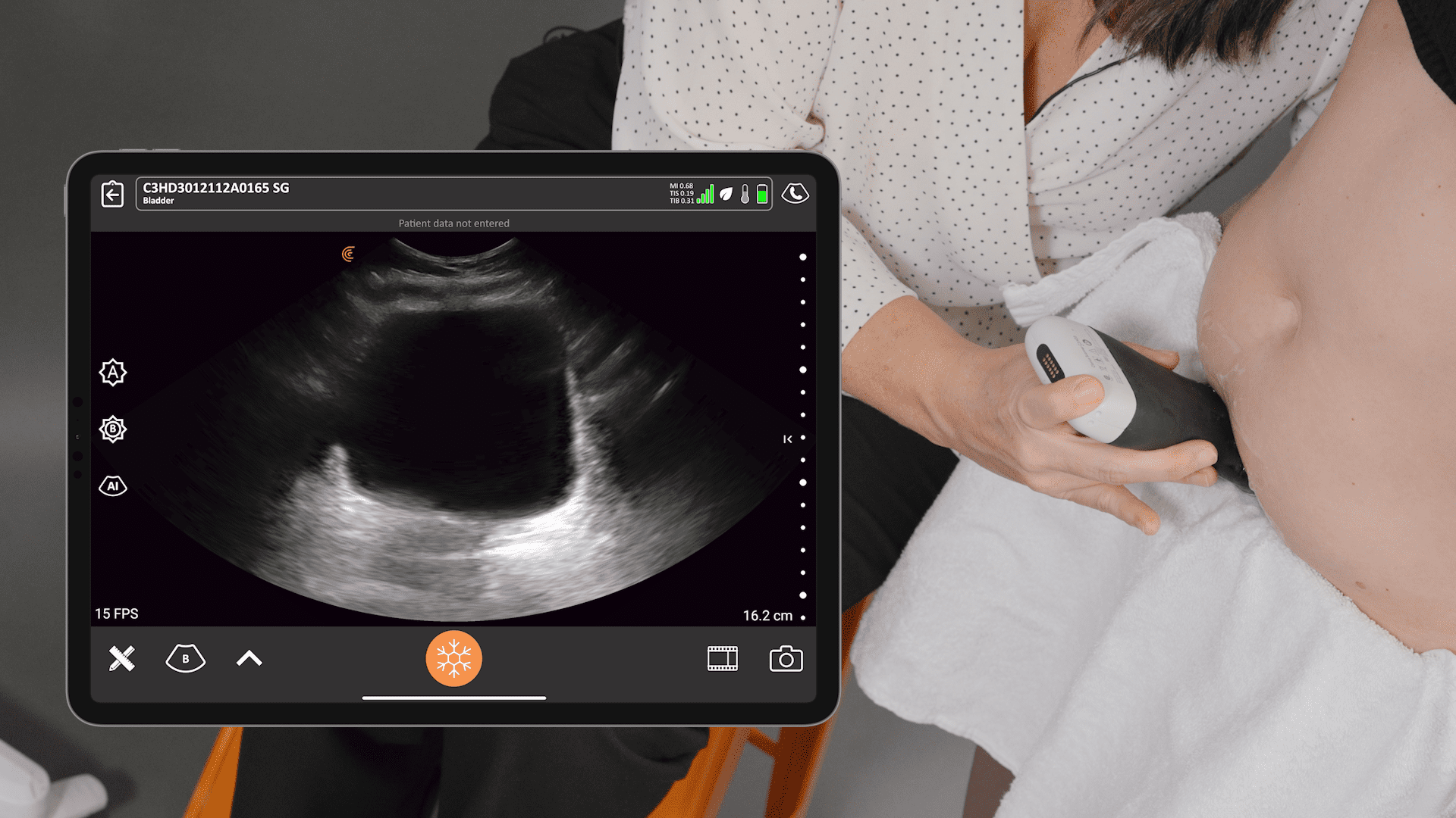
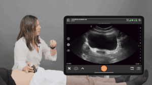
Scan In-Clinic with Clarius Ultrasound for Fast Insights on the Spot
Delivering the same high-powered imaging as the best cart-based systems, Clarius HD3 ultrasound shows you the fine details you need to quickly investigate an area of concern for an accurate diagnosis. Our linear scanners are unrivaled for high-resolution musculoskeletal imaging and procedure guidance, with an easy-to-use app powered by artificial intelligence and connected to the cloud. With AI assistance on your smart device, getting a great image is easy. Start scanning in seconds, then simply pinch to zoom, slide to change gain, and tap to switch modes.
To learn more about how easy and affordable it is to add Clarius HD3 to your practice, contact us today or request a personalized virtual ultrasound demo.


