


Aesthetic procedures, particularly those involving facial fillers, have become increasingly popular. However, they also carry potential risks and complications, which can be mitigated with the use of ultrasound guidance.
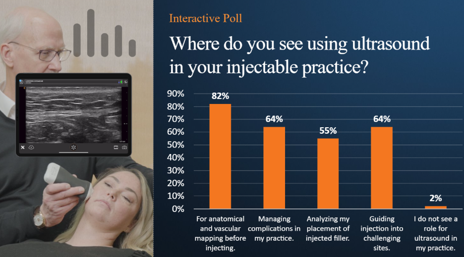
Dr. John Arlette, a dermatologist and expert in the use of ultrasound in aesthetic procedures, joined us to present a webinar on how ultrasound can be used to enhance safety and precision in jawline rejuvenation. More than 1700 clinicians registered for the popular webinar. A poll of attendees revealed that most clinicians are considering ultrasound for anatomical and vascular mapping before a procedure.
The free webinar recording is available now to watch at your convenience: Ultrasound for Safe Jawline Rejuvenation: Visualizing Anatomy, Guiding Filler Injections, and Resolving Complications. Read on for some key takeaways and a demonstration video from the webinar.
Why Ultrasound Matters in Jawline Rejuvenation
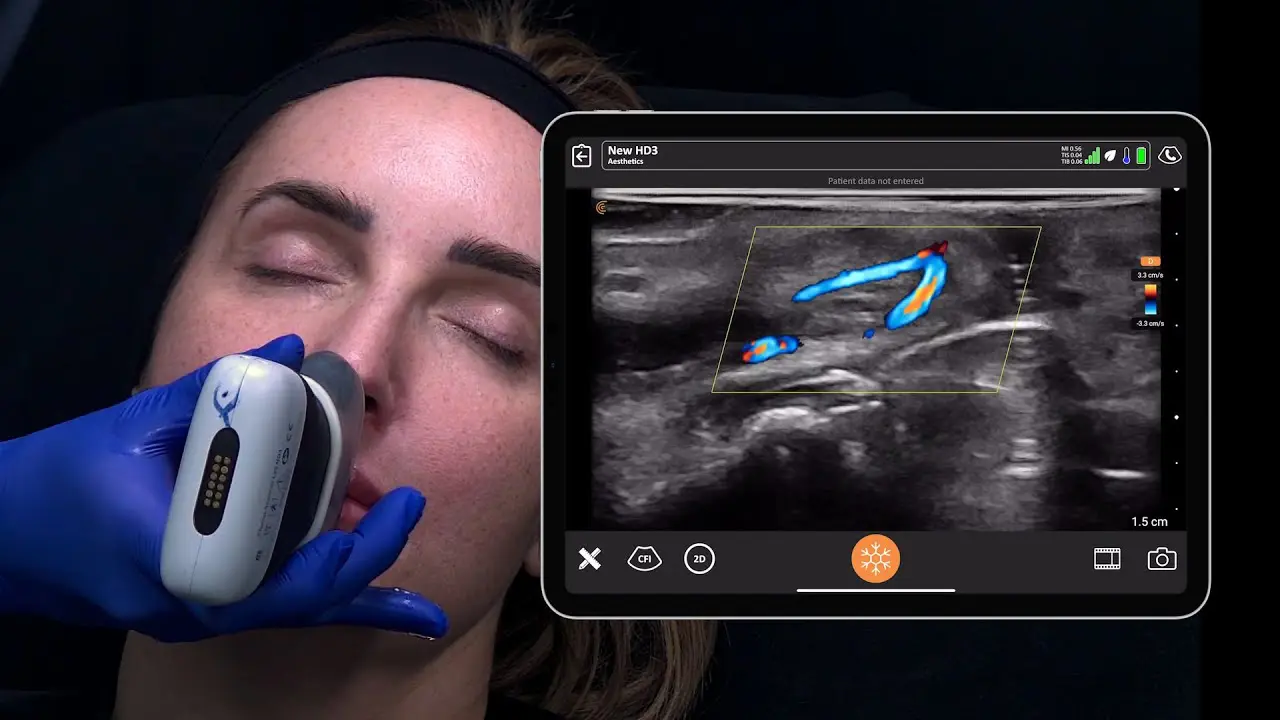
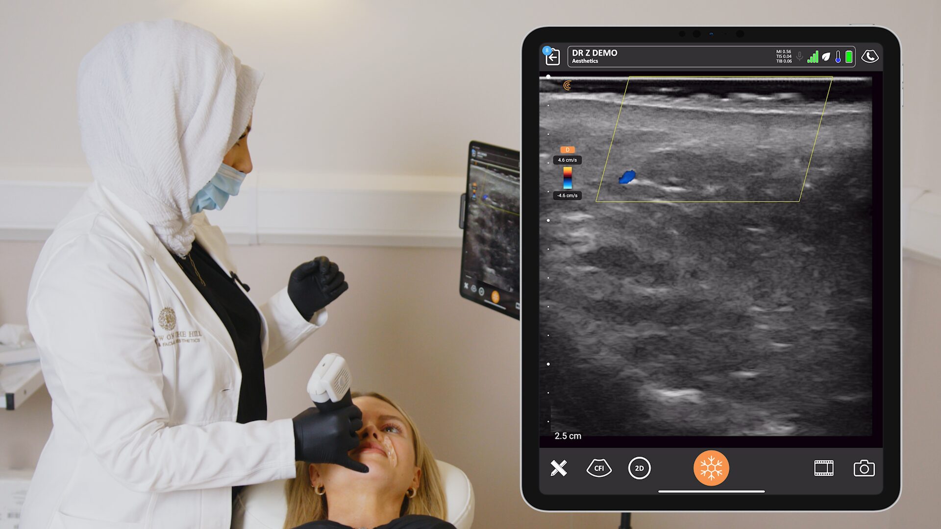
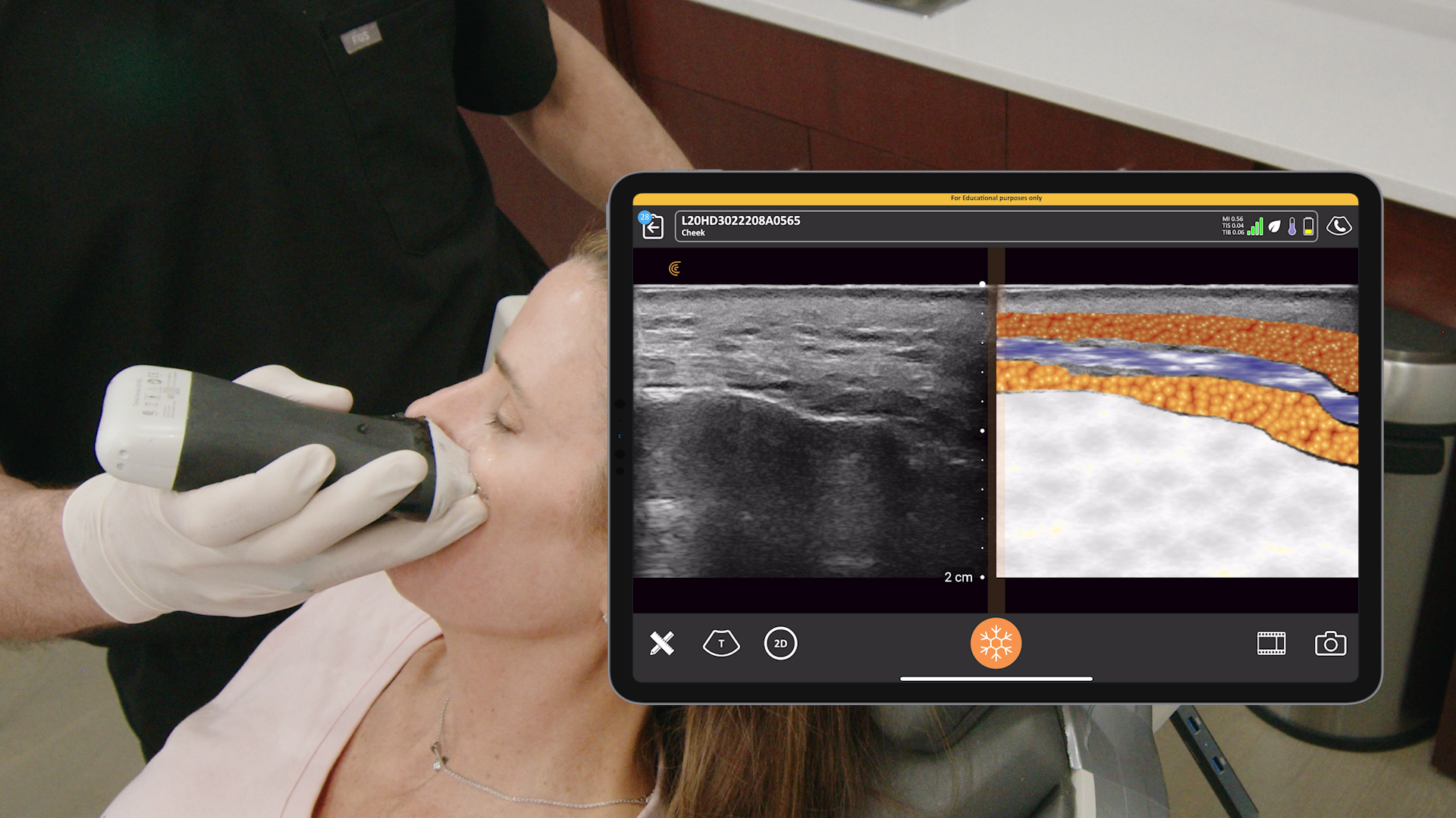
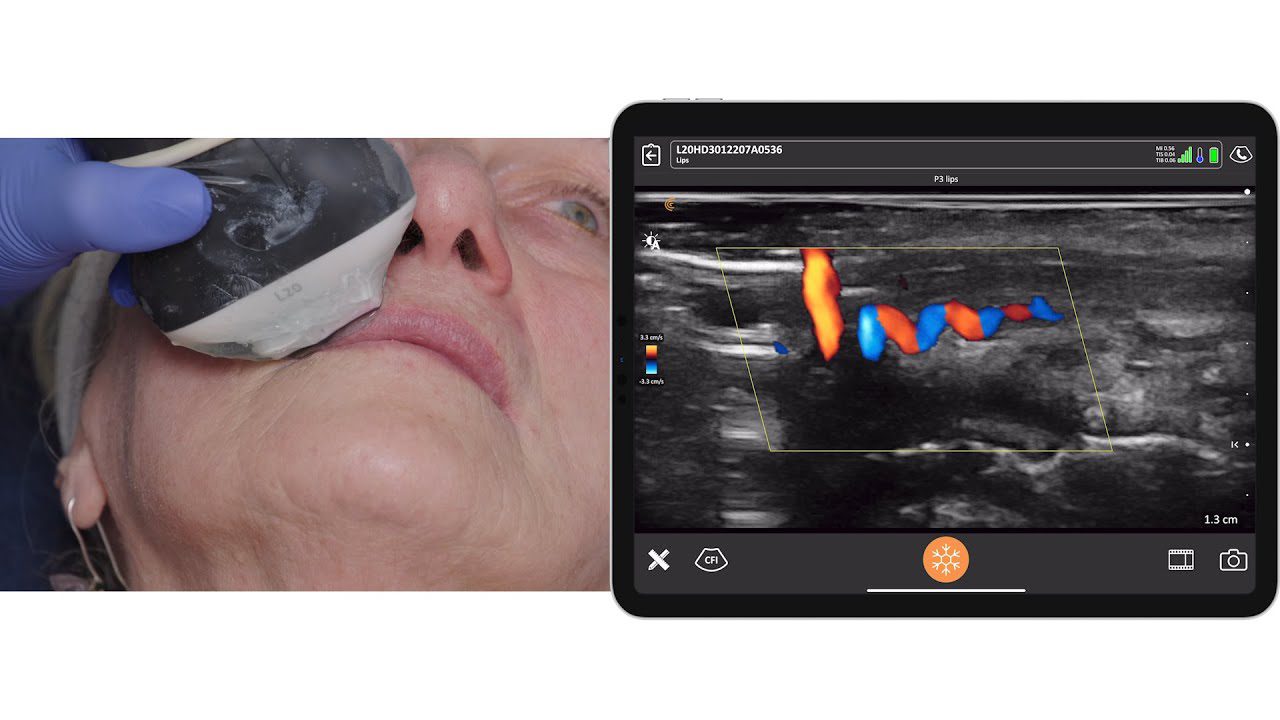
- Visualizing Anatomy: Ultrasound allows for real-time visualization of critical anatomical structures, such as blood vessels and the parotid gland, reducing the risk of complications like vascular occlusion and incorrect filler placement.
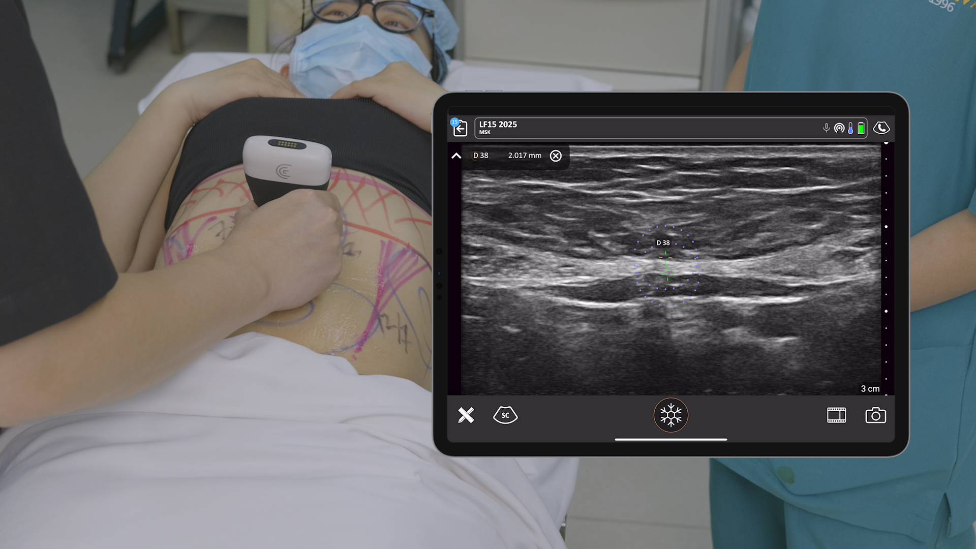
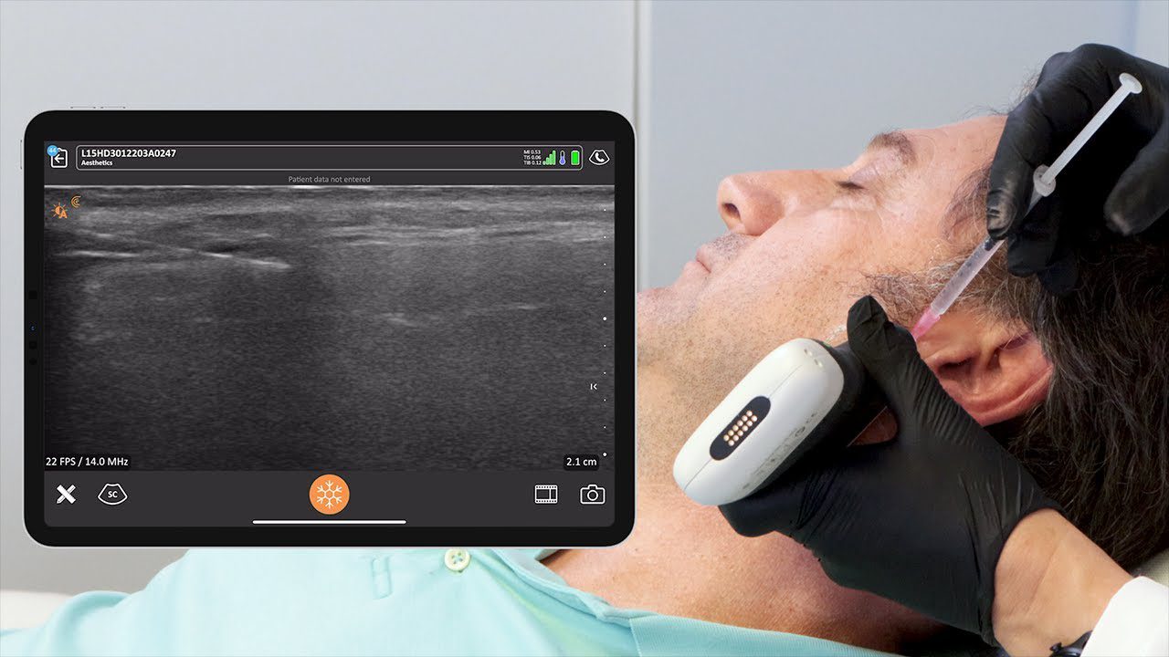
- Guiding Filler Injections: Ultrasound guidance ensures accurate filler placement, leading to more predictable and aesthetically pleasing results.
- Managing Complications: Ultrasound can be used to identify and characterize complications, such as abscesses or misplaced filler, facilitating prompt and effective management.
Key Points from the Webinar
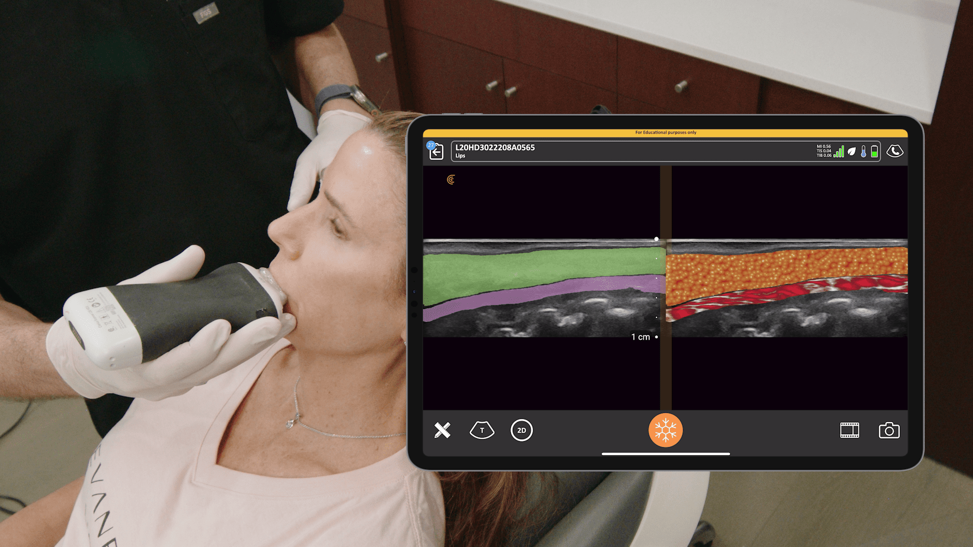
- Understanding Jawline Anatomy: The jawline is a complex area with variable anatomy. Ultrasound allows for precise identification of key structures, including the masseter muscle, parotid gland, facial artery and vein, and various facial muscles.
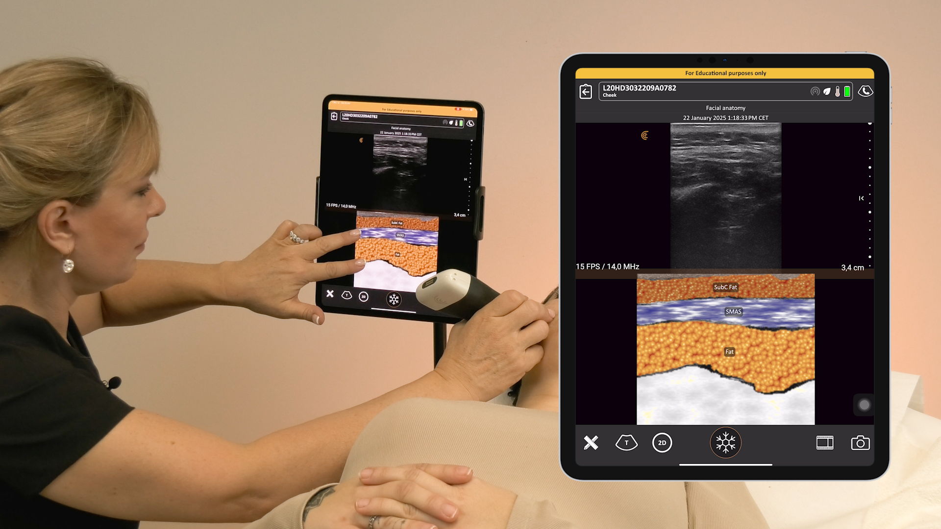
- Safe Injection Techniques: Ultrasound guidance enables injectors to avoid critical structures, ensuring safe and effective filler placement. Injecting above the SMAS (superficial musculoaponeurotic system) can help prevent nodules and achieve better lifting effects.
- Identifying and Managing Complications: Ultrasound can be used to diagnose and guide the treatment of complications, such as inflammation of the parotid gland and masseteric nodules. Aspiration and hyaluronidase injections can be performed under ultrasound guidance for optimal results.
- Benefits of Ultrasound in Practice: Ultrasound enhances patient safety, improves treatment outcomes, and increases injector confidence. It also allows for objective assessment of treatment results and can be used for patient education.
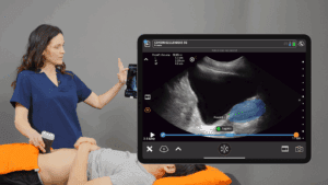
Training Builds Confidence
Integrating ultrasound into your practice can elevate the standard of care you provide and achieve optimal results for your patients. Hands-on training courses or workshops offer practical experience and confidence in using ultrasound for aesthetic procedures. Access online training webinars and videos to get you started on the basics.
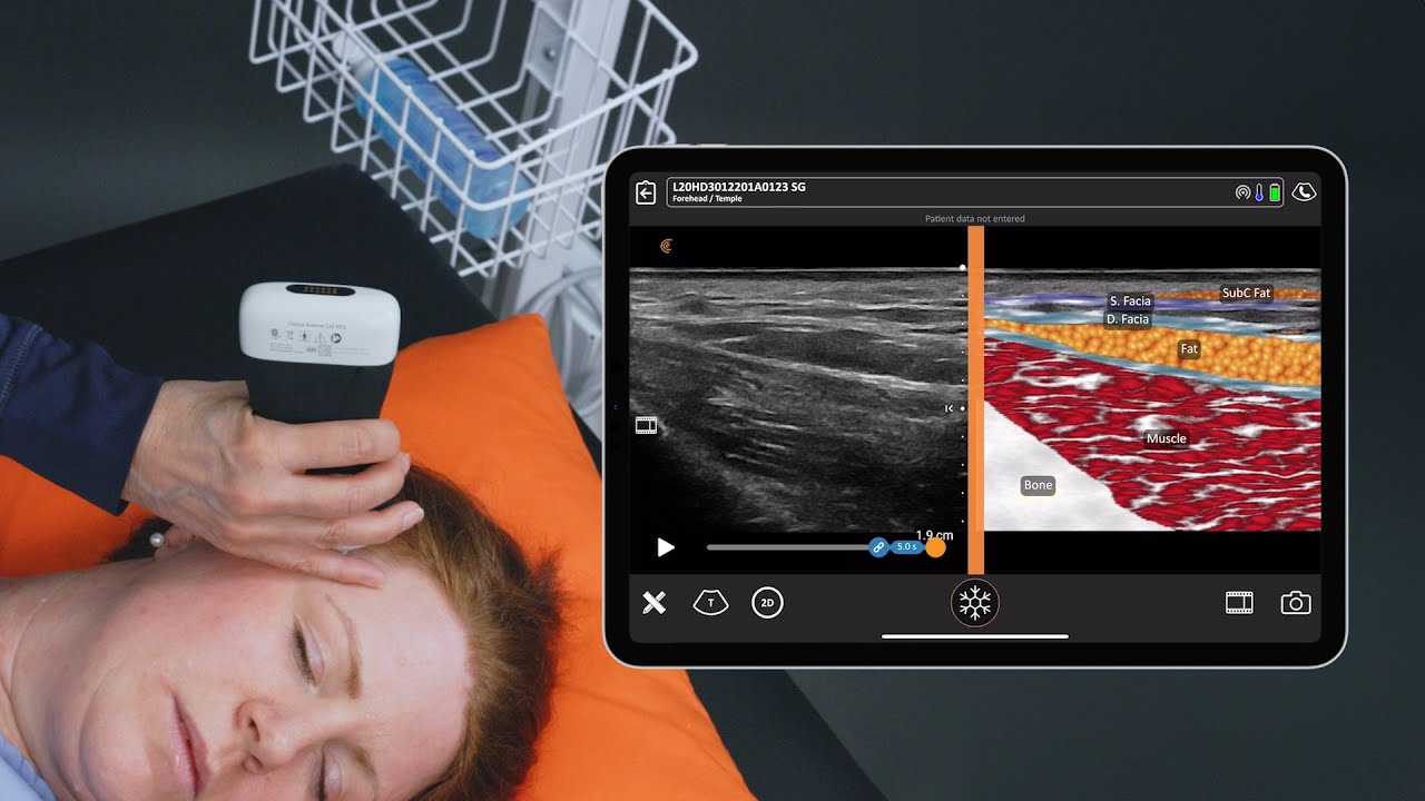
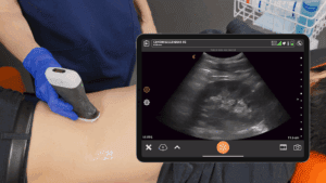
Watch this 8-minute video to see Dr. Arlette’s systematic ultrasound exam of the mandible, from the angle of the jaw to the chin, identifying structures that are relevant and important for successful filler or neurotoxin injections. He uses the Clarius L20 HD3 handheld ultrasound scanner.
AI-Powered Clarius Handheld Ultrasound: The Leading Choice for Facial Aesthetics
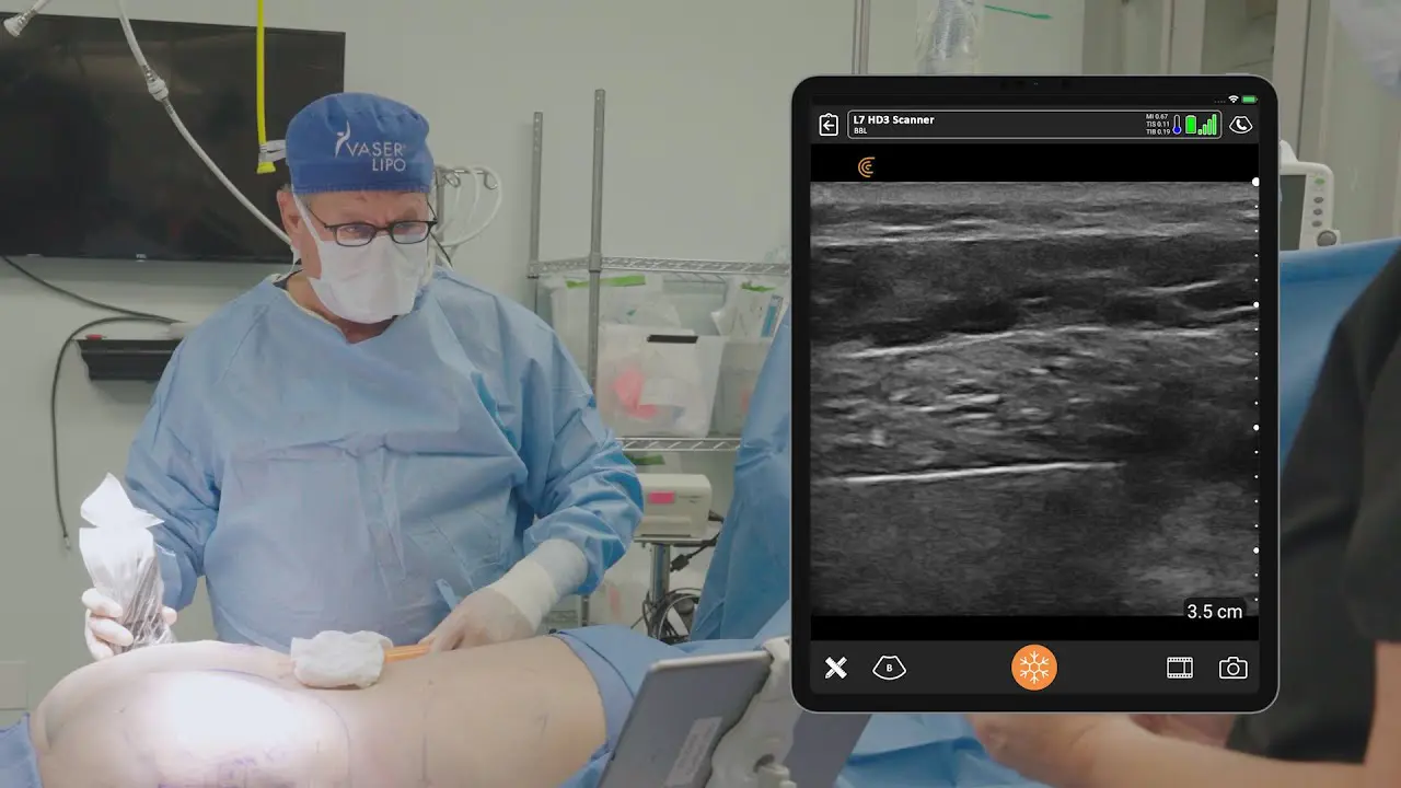
Wireless and pocket-sized, Clarius handheld ultrasound scanners deliver the high-definition imaging and performance of traditional ultrasound systems for a small fraction of the cost. They are the leading choice for plastic surgeons and aesthetic practitioners performing ultrasound-guided procedures to ensure patient safety.
New T-Mode™ by Clarius is a groundbreaking educational technology to help clinicians new to ultrasound advance their image interpretation skills using Clarius handheld scanners.
Visit our aesthetics page to learn more or request a virtual ultrasound demo today to learn how high-definition ultrasound imaging with voice controls can improve safety and deliver consistent patient outcomes at your aesthetic practice!
