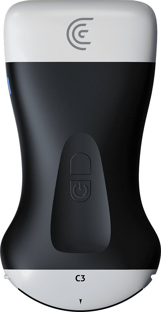
hi I'm Dr Kevin Zorn and today I'll be performing a clarius ultrasound for my patient looking at his bladder and his prostate to evaluate his lower urinary tract symptoms so I have the C3 the small curved array third generation Clary system and I'll be linking up now to my Claris app on my iPhone to have the pre-programmed prostate settings so it'll best discriminate the tissues around the bladder and the prostate so let's begin so I've already applied the gel to the patient's lower abdomen just above the pubic bone and I'll apply and start the filming so up front so I can see clearly his bladder I'm going to scan and fan from the upper part of the Dome of the bladder and scan downward nice and slowly getting an idea if there's any bladder wall defects diverticulum and if there's any filling defects or hyperechoic areas suggestive of a bladder stone so here we go scanning up and down clearly a nice homogeneous a Smooth Wall of the bladder and I'll usually pick the largest aspects of this bladder which looks pretty full and I'll go ahead and measure the bladder wall thickness so typically this gives us an idea if there's some hypertrophy of the prostate which will create more resistance of a bladder Outlet obstruction and create some thickening of the bladder wall so we'll go ahead and save that image moving my way down at the bladder neck to see if there's any median lobe any protrusion essentially of the prostate into the bladder so here we can see The prostated View I can zoom in centralize the image and there you go so I don't see any medial lobe if you want we'll just go sagely do the same views again just making sure that there's no anterior wall tumor or defect so clearly Fanning from both sides of the bladder clearly no abnormalities so I'll go ahead and freeze sack for that image that's my third of the bladder then I'll come back to the Midview of the bladder neck and this begins my Contour of the prostate anatomy so this is where I'll depress down on the belly you have certain patients who are more obese we'll ask them to lift up their penis so that it flattens the area I usually want to push downward and cephalad toward the head and then get underneath the pubic bone to avoid the ultrasound being bounced off the pubic bone which can get in the way for a view of the Apex so here we go clearly small prostate so I'm going to Fan it out and I can zoom in so they're the seminal vesicles of the little bunny ears behind the prostate those are normal and as I fan downward there's our prostate just getting an idea of the echogenicity is there any hypoechoic areas or are there calcifications suggestive of inflammation or chronic inflammation and let's go ahead and save that large view and do my prostate volume measures so there's my upper my lower scoot that up and then same thing for the width save that so you can notice I'm doing this all with my iPhone with my thumb so this is all readily available quick to your finger and rapidly give your findings so again same idea torque down I'm looking for the seminal vesicles here they are one and two and take that largest View of the prostate here we go we can see the funneling of the bladder neck and measure its to its width and I'll save that so there all within a matter of a few minutes I've got the pertinent information for anyone with BPH or lower urinary tract symptoms where we will have their bladder status their bladder wall thickness making sure there's no secondary endpoints of chronic BPH just as diverticulum or stones and finally the prostate anatomy length height and width and the absence or presence of a median lobe so this completes the ultrasound findings and I'm going to go ahead now and move toward a final summary report so I'll be able to enter in the patient's information and generate really smoothly with a click of a button that some of the findings indications here was BPH there's no catheter in place who the referring physician was and simply scrolling down you can see the Impressions pre-modeled into the app for the prostate we can actually see it was normal there's no other hyperchoric or calcifications areas it was symmetric the seven of us as we saw there was no medial lobe the bladder looked normal at that point I can either use Siri dictate or type in my recommendations and notes and once complete sent to the cloud for implementation into the patient's chart or to the refrain position so that completes our clarius C3 evaluation of the prostate and bladder for this gentleman foreign