Dr. Oron Frenkel

Classification of a mass with vascular flow can limit or expand the differential diagnosis, in this case creating suspicion for lymphoma.
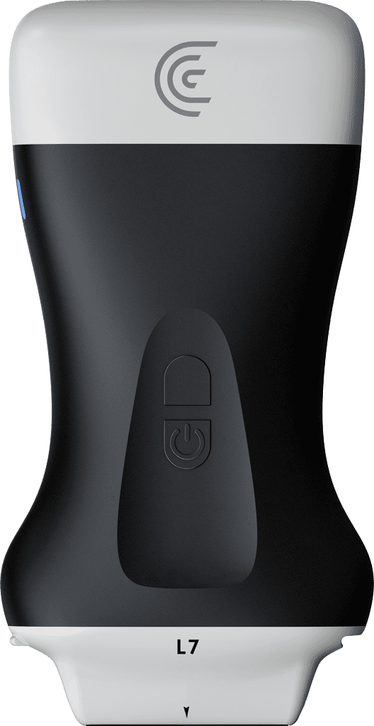
Specialties: Emergency Medicine, Pediatrics, Primary Care, Rural
Applications: Superficial
in this video we see the linear scanner on a soft tissue preset placed in the supraclavicular fossa in a patient with a mass that was identified the mass can be clearly seen here a budding and artery likely the subclavian artery and the mass has a very well demarcated boundary and there was a question of whether this was infectious or inflammatory there is some echogenic material inside the mass suggestive that is not fluid filled and then placing power doppler on identifies clearly there is vascular flow within the mass making it highly suspicious for a lymph node or some other kind of malignancy in this case it was highly suspicious for lymphoma given the location in the supraclavicular fossa

Products Used

More Classes

Request a Quote

With 8 scanners available, we offer a wireless ultrasound solution tailored to your needs. Request a quote to discover which scanner can deliver the best ultrasound imaging for your practice.

⚠️ Clarius ultrasound is for medical professionals only.

By providing my email, I consent to receive Clarius webinar invitations, case studies, whitepapers, and more, and I consent to the Clarius Privacy Policy. I can unsubscribe anytime.