



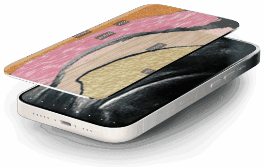

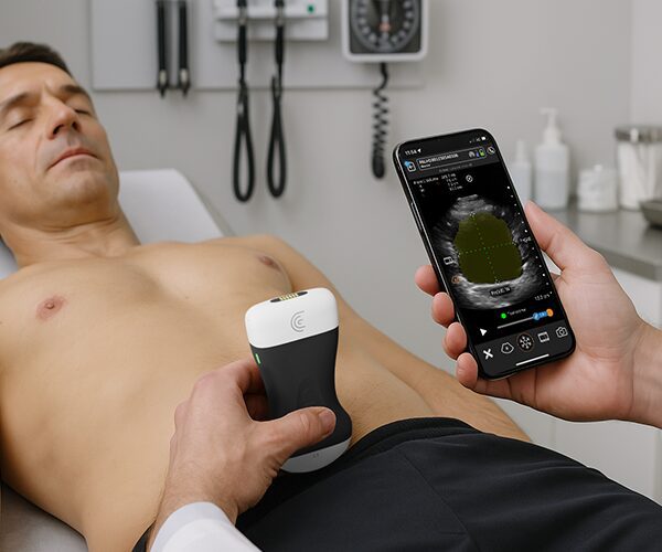


Panoramic imaging (extended field of view) allows clinicians to capture full-length structures like long muscles and tendons, that exceed the standard footprint of a B-mode ultrasound probe. In this step-by-step tutorial, learn how Clarius Panorama stitches a single linear sweep into a seamless, high-resolution composite image right on your device.
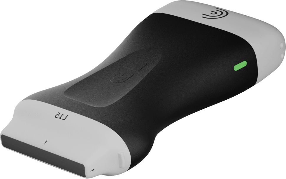
Using any Clarius linear, in this case the L7, and the patellar tendon as a clinical example, see how to transition from live scanning to full composite visualization, annotation, and measurement.
Step-by-Step Workflow Covered:
Setup: Select your scanner (e.g., Clarius L7) and appropriate clinical preset (e.g., Knee preset).
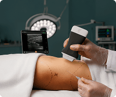
Activation: Position the probe at the target starting point, open the Modes menu, and select Panorama.
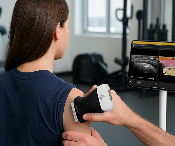
Sweeping: Execute a smooth, steady sweep along the linear path using the live reference window as your guide.
Composite View: Press Freeze to display the stitched panoramic image.
Measurement & Storage: Add annotations, measure total structure length, and save composite scans directly to the Clarius Cloud.
Key Clinical Applications:
MSK Ultrasound: Evaluating entire muscle bellies, long tendon structures (like the patellar or Achilles tendon), soft tissue lesions, and more.

