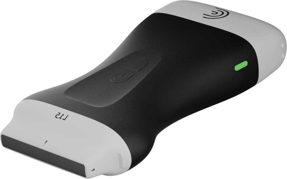
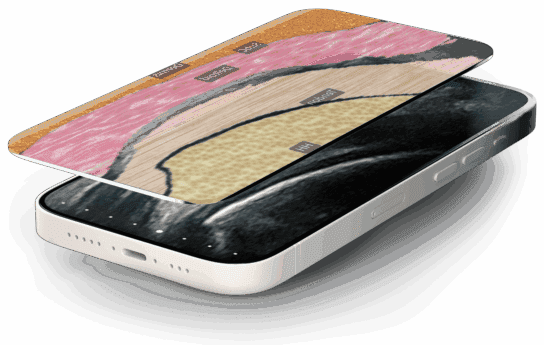
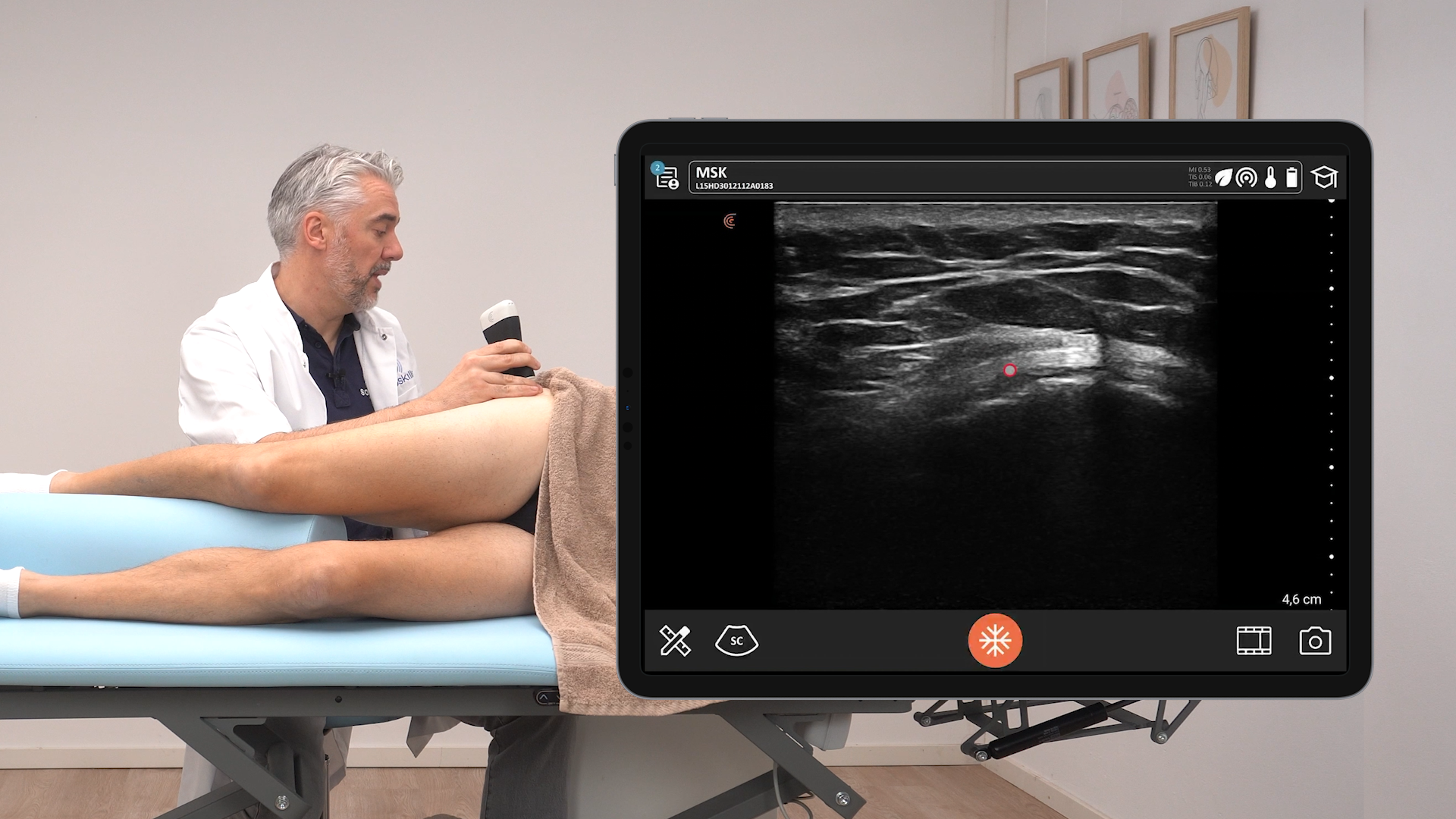
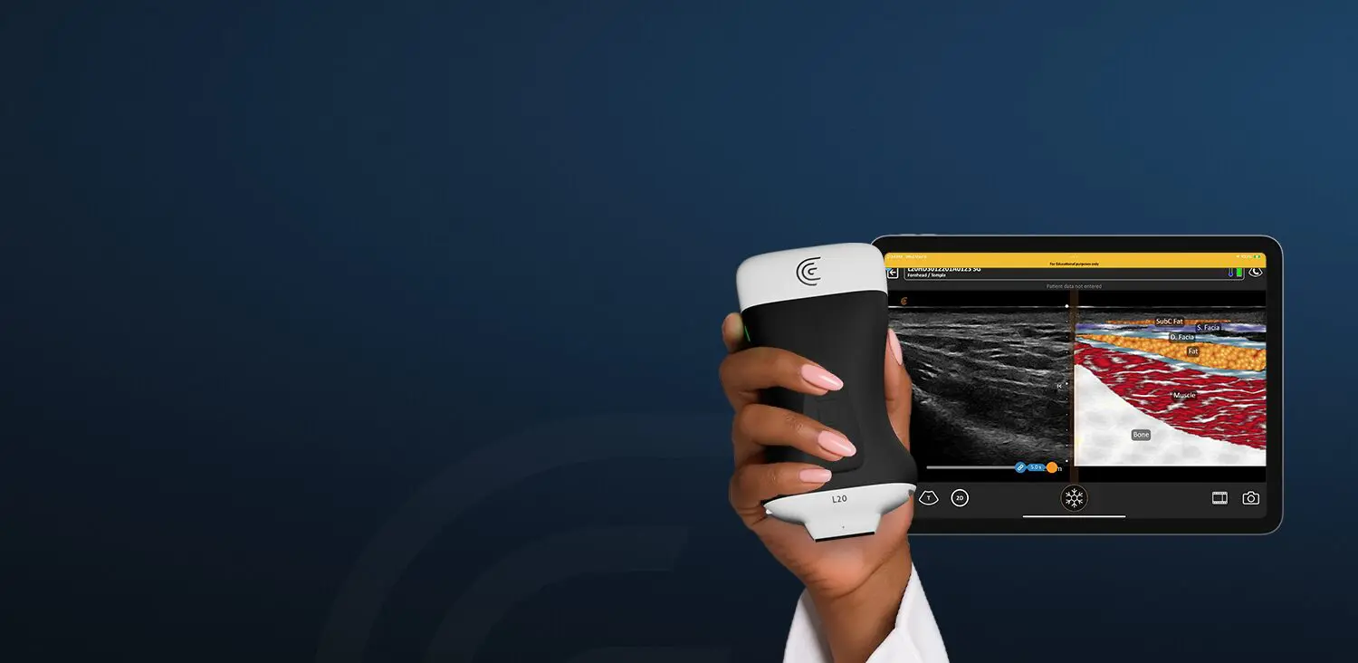
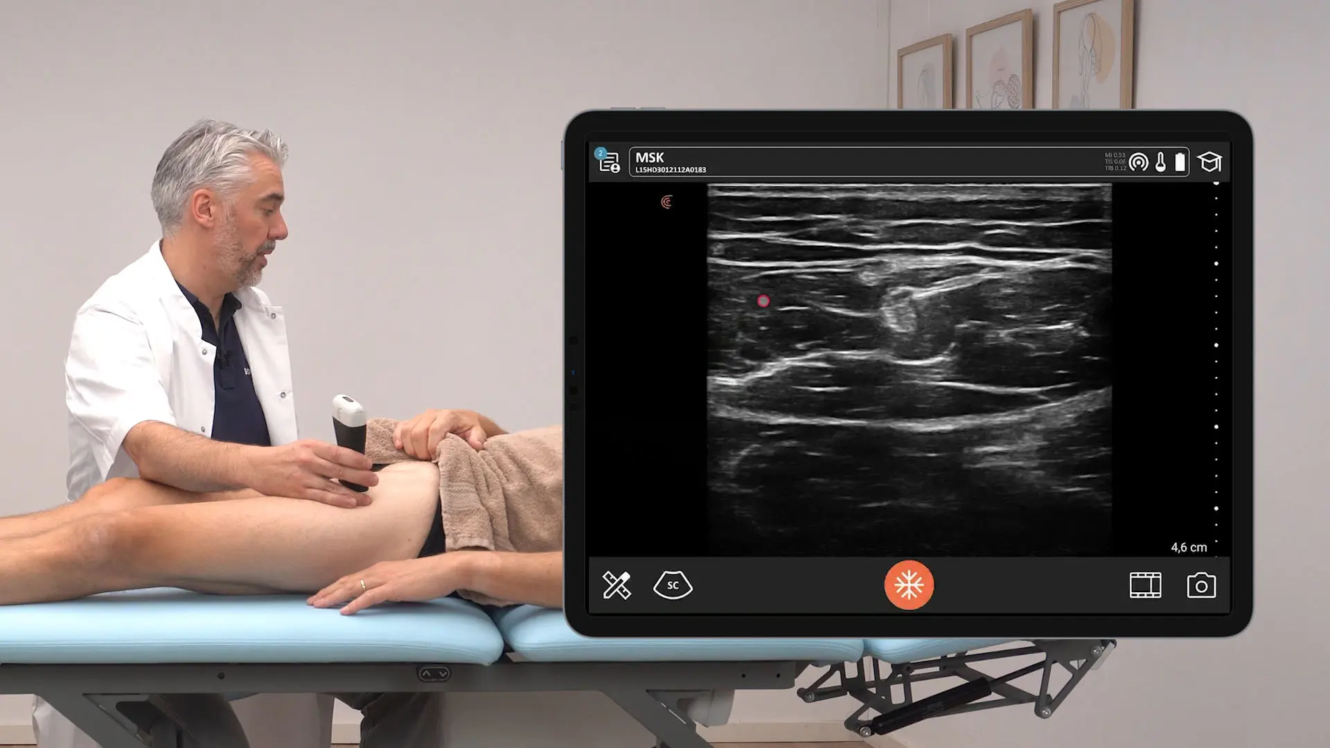
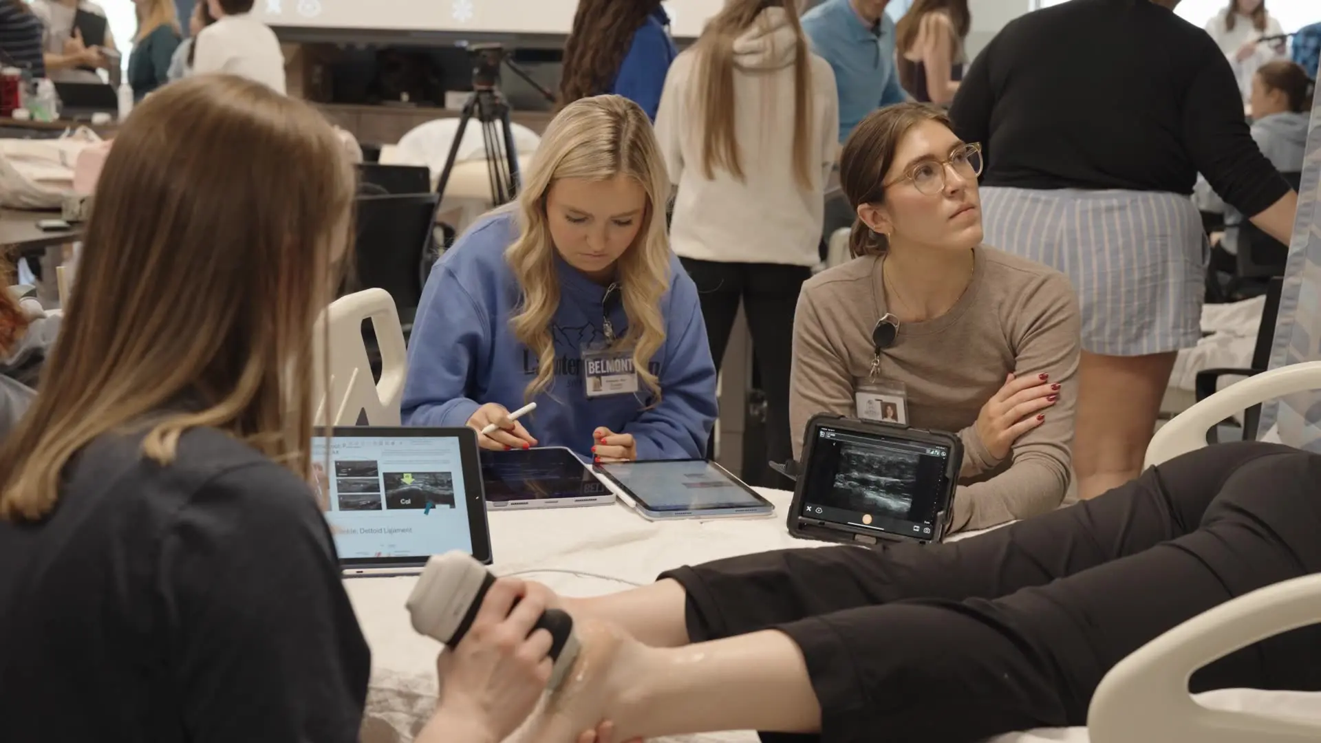
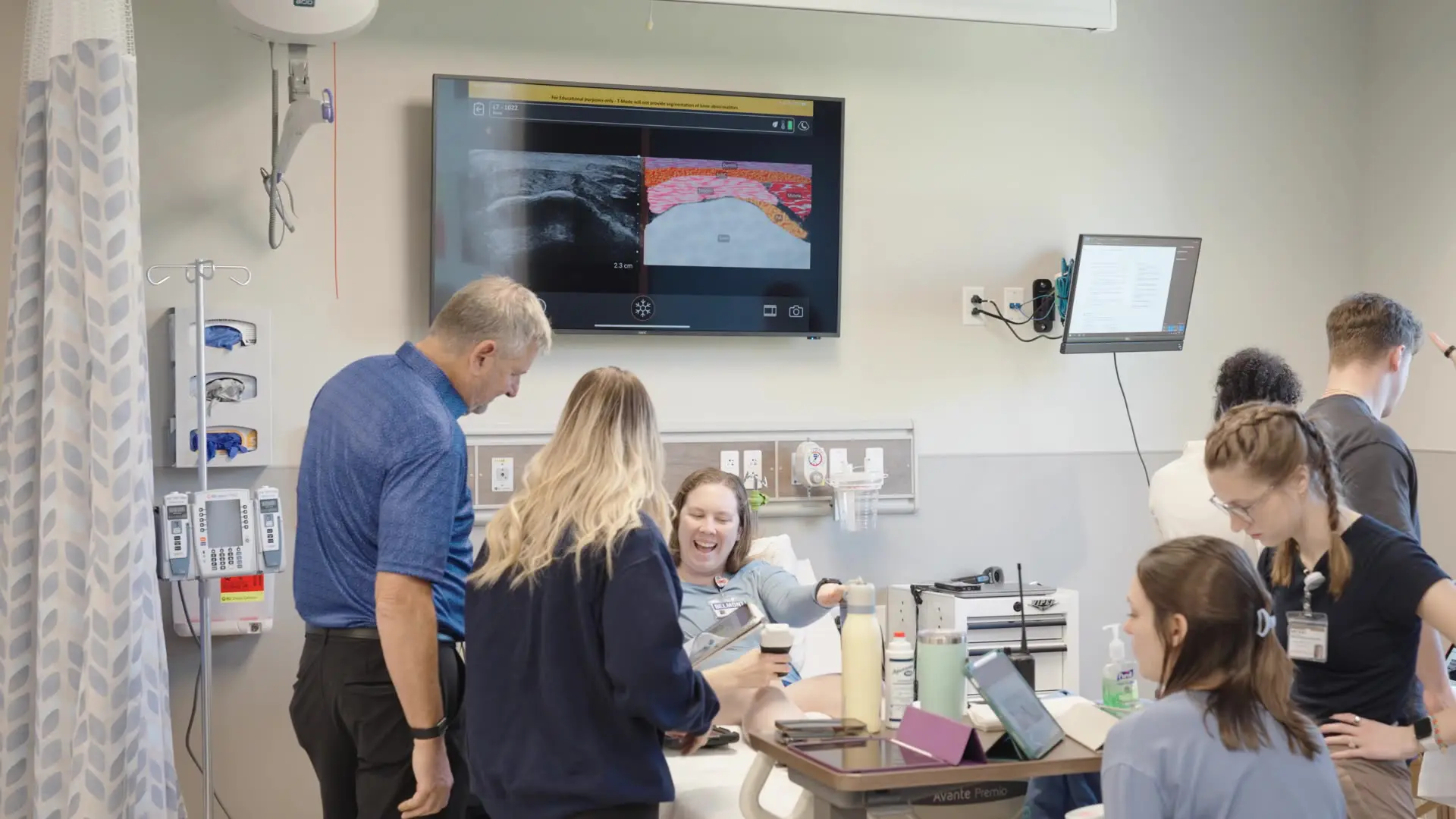
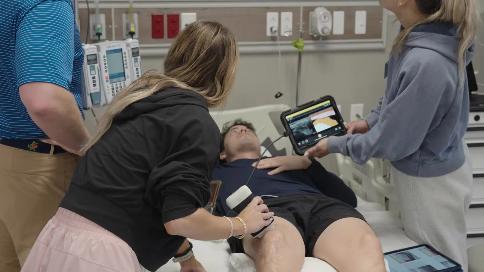
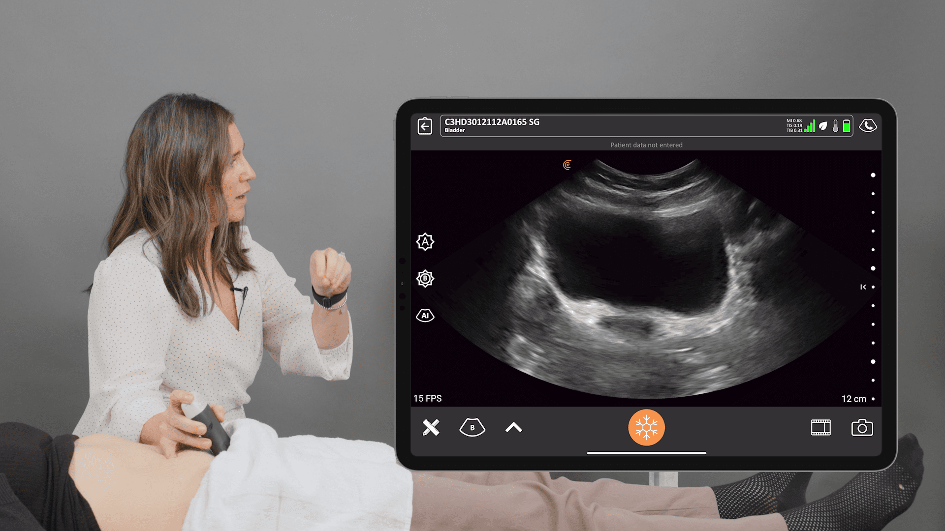
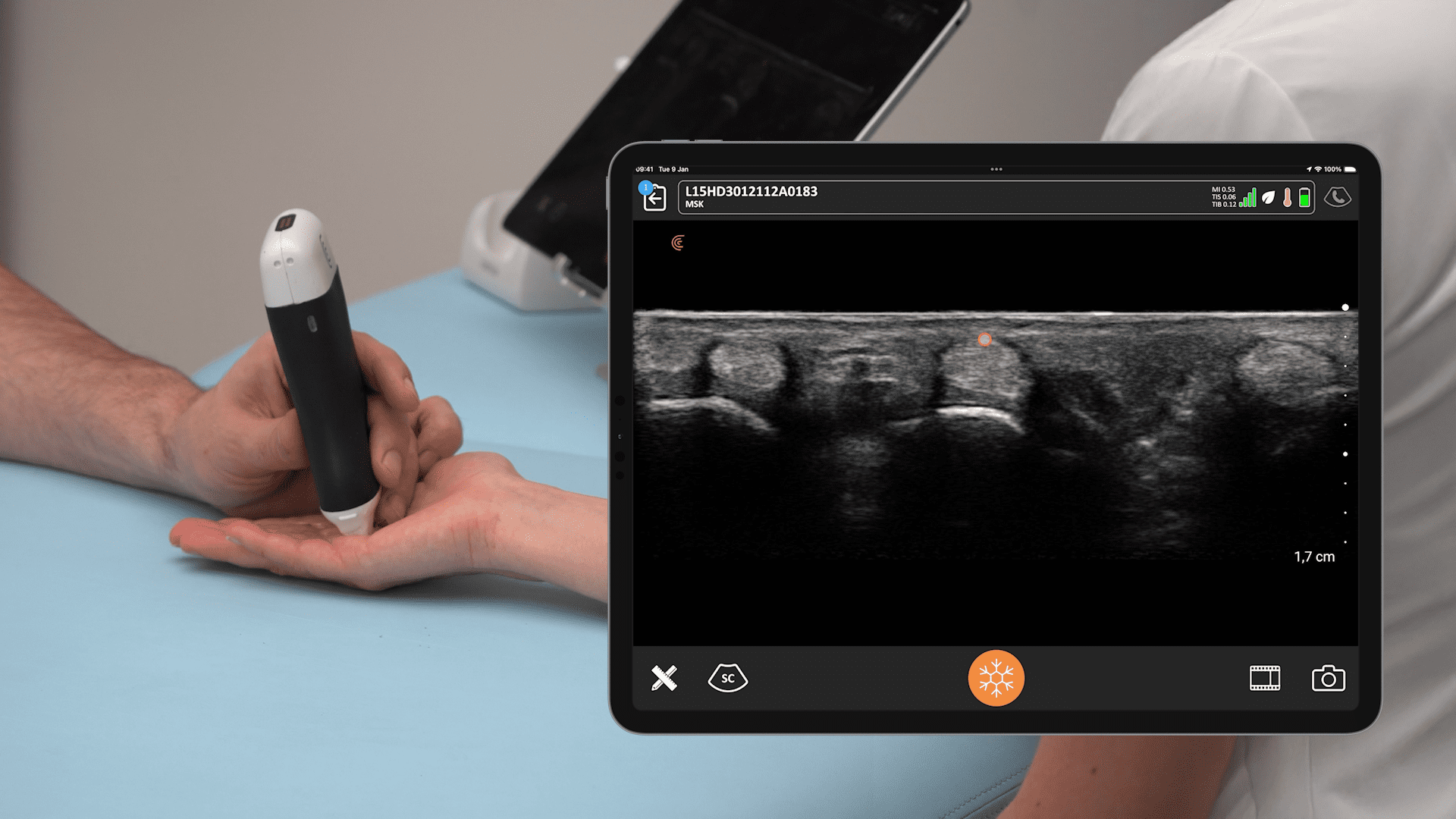
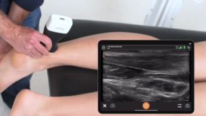
As ultrasound systems become smaller, more affordable and easier to use, we’re seeing growing interest in our handheld systems from clinicians treating musculoskeletal (MSK) ailments.
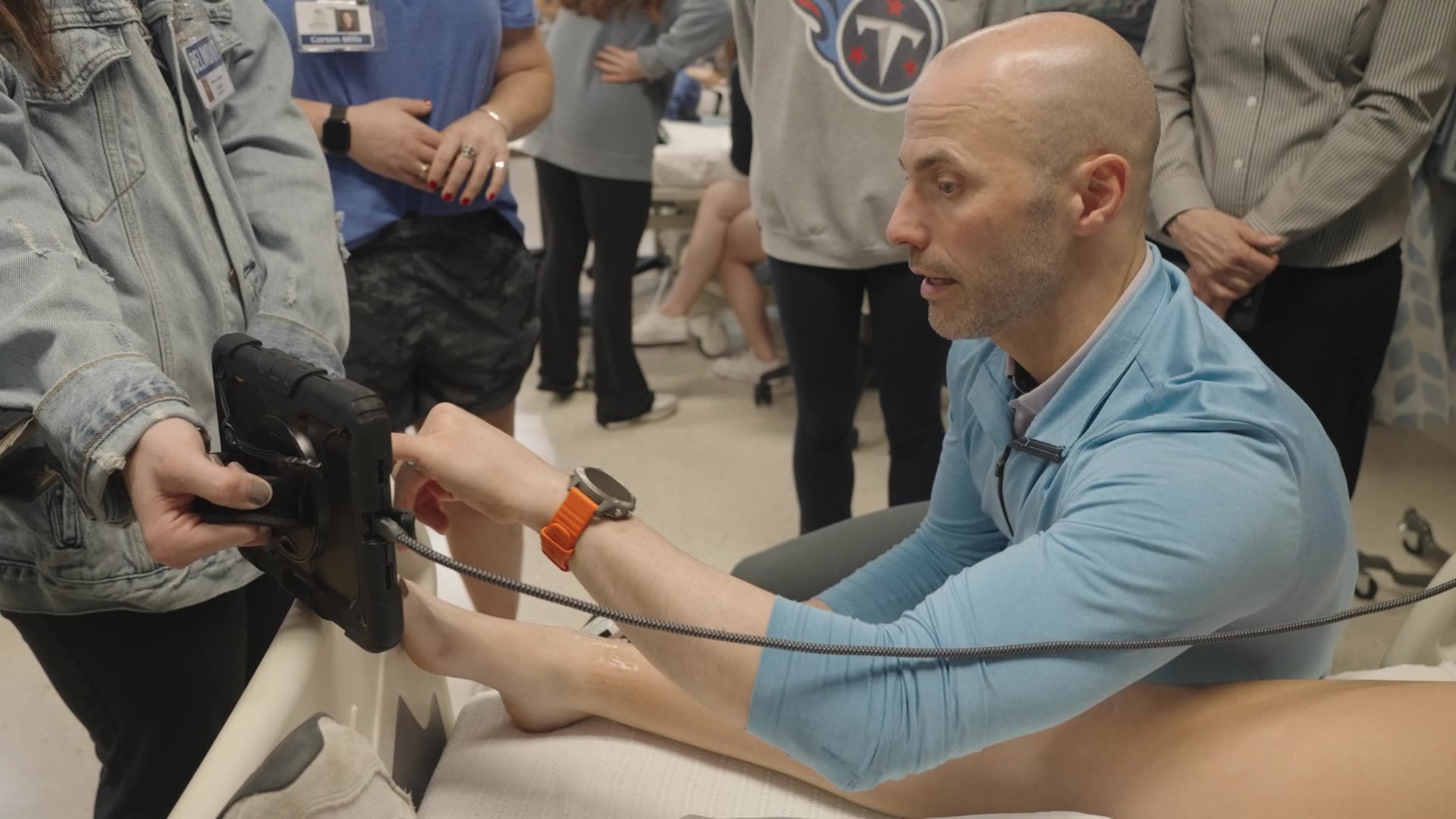
For MSK complaints, there can be so many possible diagnoses,” says MSK sonologist Greg Fritz, PT, DPT, RMSK. “We know from many studies that ultrasound is superior for diagnostics than a physical exam. Being able to see the issue in real-time means I don’t have to order x-rays or wait for the results from an MRI. It saves the patient pain and time.”
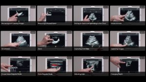
We worked with Dr. Oron Frenkel, MD to create a series of brief instructional videos on MSK ultrasound using the Clarius L15HD high-frequency linear ultrasound scanner. Watch the videos below or in the Clarius Ultrasound App!
How to Scan the Elbow Using Clarius Handheld Ultrasound
A survey ultrasound scan of the elbow can identify some of the most common pathologies that cause acute and chronic elbow pain such as tendinosis of the common flexor or extensor tendons, joint effusion or olecranon bursitis. Watch this video to learn how to perform a focused elbow scan using ultrasound. Dr. Oron Frenkel uses the Clarius L15HD to look for signs of disruption, calcification and fluid on the tendon.
Ultrasound Exam of Olecranon Bursitis
In this 1-minute video of an ultrasound exam of an elbow, you’ll see a fluid-filled bursa superficial to the olecranon.
How to Scan the Shoulder Using Clarius Handheld Ultrasound
An overview ultrasound scan of the shoulder can identify some of the most common pathologies that cause acute and chronic shoulder pain such as rotator cuff tendinopathy and tears, joint effusion and bursitis. Watch this video to learn how to perform a dynamic shoulder exam. Dr. Oron Frenkel uses the Clarius L15HD with the shoulder preset to look at for full-thickness tears or calcification.
Ultrasound Exam of a Rotator Cuff Tear – Short Axis
Unlike the previous video, which shows what a normal rotator cuff looks like using ultrasound, in this 30-second video, you see Dr. Oron Frenkel discover discontinuity of the rotator cuff, which suggests a complete rotator cuff tear.
Ultrasound Exam of a Rotator Cuff Tear – Long Axis
Watch this video to see what a rotator cuff tear looks like during an ultrasound scan in long axis.
Ultrasound Exam of Shoulder Impingement
In this 50-second video captured during dynamic testing while the patient abducts, you’ll see a positive impingement sign when the humerus is seen striking the acromion in real-time, which suggests significant rotator cuff pathology.
Physical Therapy Case: Rotator Cuff Injury
During a recent webinar, Dr. Greg Fritz presented the following case of a real patient who benefitted from MSK ultrasound. Edwin is a family practice physician who injured his shoulder while moving boxes in his new home. He has pain when lifting his arm and intermittent lateral shoulder and forearm numbness. He came to Dr. Fritz to see how badly he tore his rotator cuff before seeing a surgeon. In this video, Dr. Fritz explains his process for conducting a shoulder cuff exam using ultrasound.
Handheld Ultrasound is Accessible for Every Clinician
Handheld ultrasound has made high-definition imaging more affordable and easier to use than complex cart-based and laptop systems. Ideal for MSK anatomy down to 7 cm, the Clarius L15 HD high-frequency ultrasound scanner is available with an optional Advanced MSK Package for diagnostic and interventional procedures. Learn more about which Clarius Handheld Ultrasound scanner is right for your MSK Practice.
You’re also invited to join renowned SonoSkills educator Marc Schmitz as he teaches anatomy, sonoanatomy, technical scanning skills and pathologies common to the shoulder, elbow and knee in the free upcoming webinar «Pragmatic MSK Ultrasound: Scanning the Rotator Interval, Common Extensor Tendon and Patellar Tendon.»
Clarius offers four ultrasound scanners that are suitable for MSK applications. Learn more about which Clarius Handheld Ultrasound scanner is right for your practice. Or contact us today to request an ultrasound demo.
