


During a recent webinar, physical therapy educators Dr. Mike Voight and Dr. Chris Wolfe of Belmont University discussed their innovative approach to incorporating Clarius wireless ultrasound into their physical therapy curriculum. They provided a behind-the-scenes look at how the university is equipping its students with essential ultrasound skills from the very start of their training. The speakers highlighted the evolving landscape of physical therapy and the increasing importance of diagnostic skills for new graduates.
The free one-hour webinar is now available to watch on demand: Reimagining MSK Education: Teaching Ultrasound for the Next Generation of Clinicians. Scroll down for key takeaways.
Closing the Training Gap and Overcoming Barriers in Musculoskeletal Ultrasound Education
Dr. Voight opened the discussion by addressing the significant training gap in musculoskeletal (MSK) ultrasound education in medical and physical therapy programs globally. Citing a recent study, he noted that while 70% of physical therapy programs surveyed said they had ultrasound education, more than 90% of those programs offered less than ten hours of training on the subject.
The primary barriers identified by a poll of webinar attendees—and echoed by the speakers—were a lack of qualified instructors and limited access to equipment. However, Dr. Voight was optimistic, stating that these barriers are diminishing due to advancements in technology and cost-effective handheld devices. He also announced a new initiative by a group of individuals working with the Ortho Academy, which is the largest academy within the American Physical Therapy Association (APTA), to develop a free, open-source curricular blueprint to help educators incorporate ultrasound into their programs.
Belmont’s Blended Training Approach
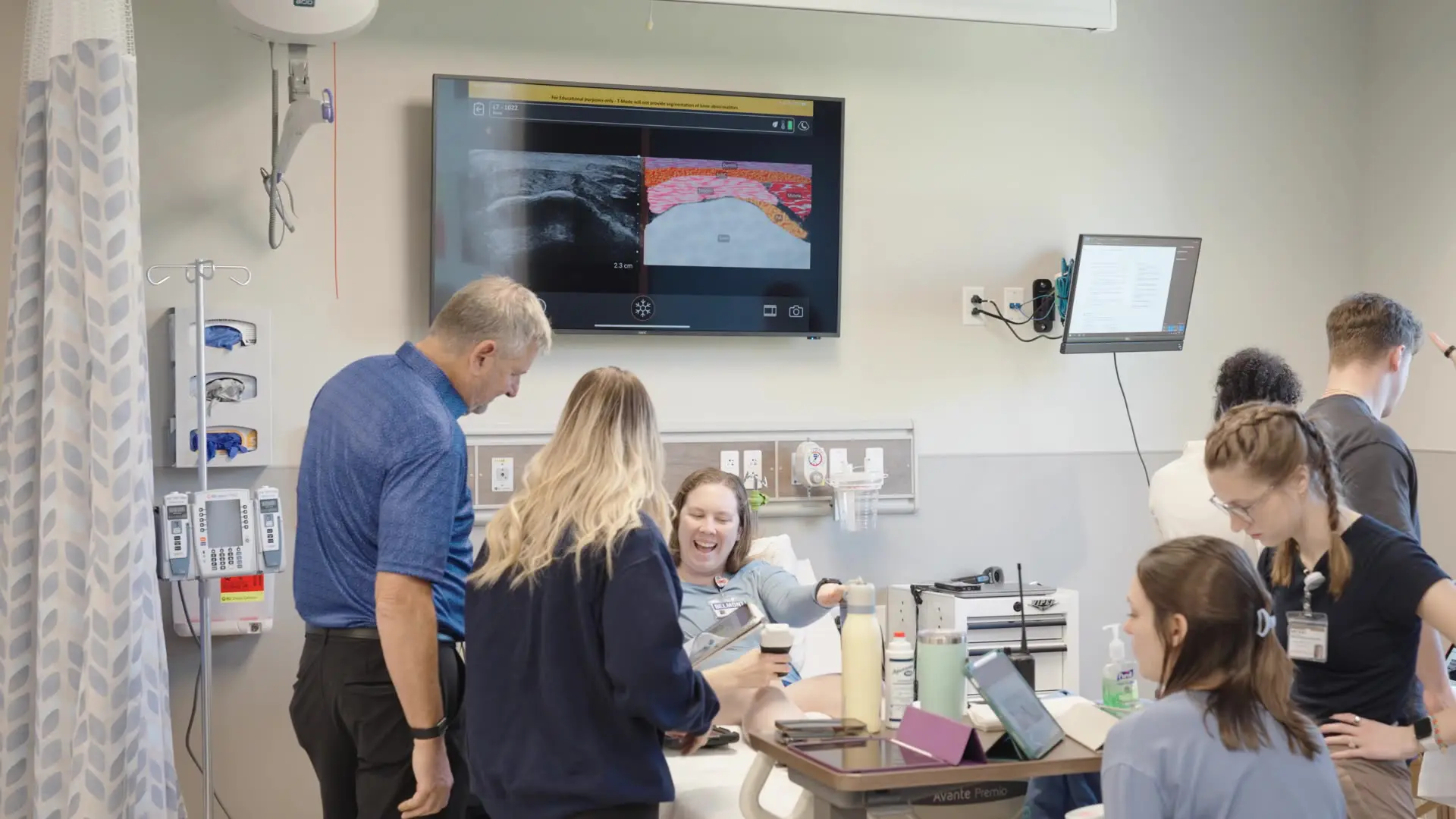
At Belmont University, Dr. Voight and Dr. Wolfe have integrated MSK ultrasound training into their three-year program’s second-year orthopedic curriculum, dedicating approximately 30 hours to hands-on learning. They emphasize that ultrasound training is not taught in isolation but is seamlessly combined with traditional examination and assessment skills.
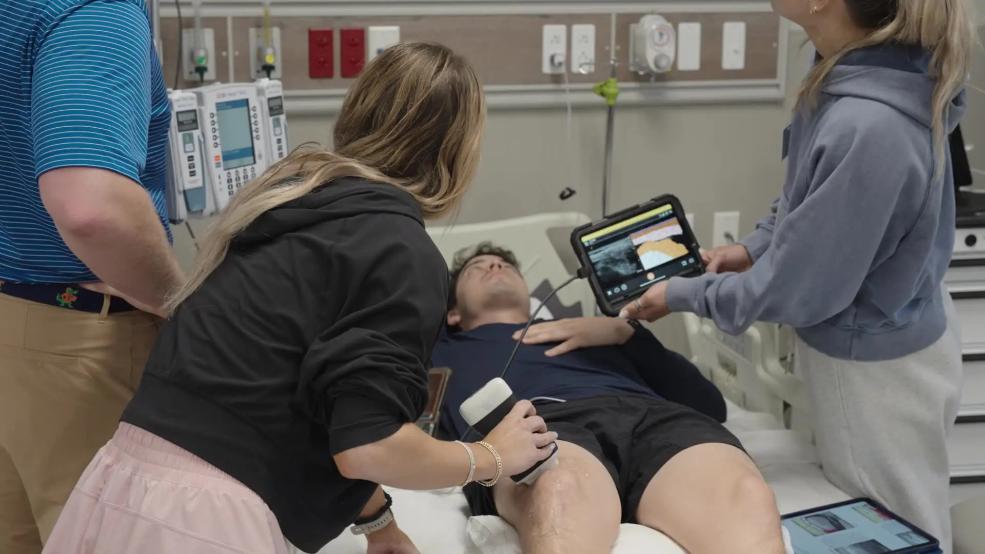
The curriculum uses a blended learning model that combines didactic lectures with active, hands-on practical sessions. Students work in groups, engaging in peer teaching and learning to differentiate between healthy and pathological tissues. Dr. Voight noted that students are surprisingly quick to grasp the process, which he attributes to its direct application of anatomical knowledge.
Watch this 3-minute video to hear what the students have to say about the hands-on ultrasound training program.
AI-Powered Ultrasound Accelerates Learning
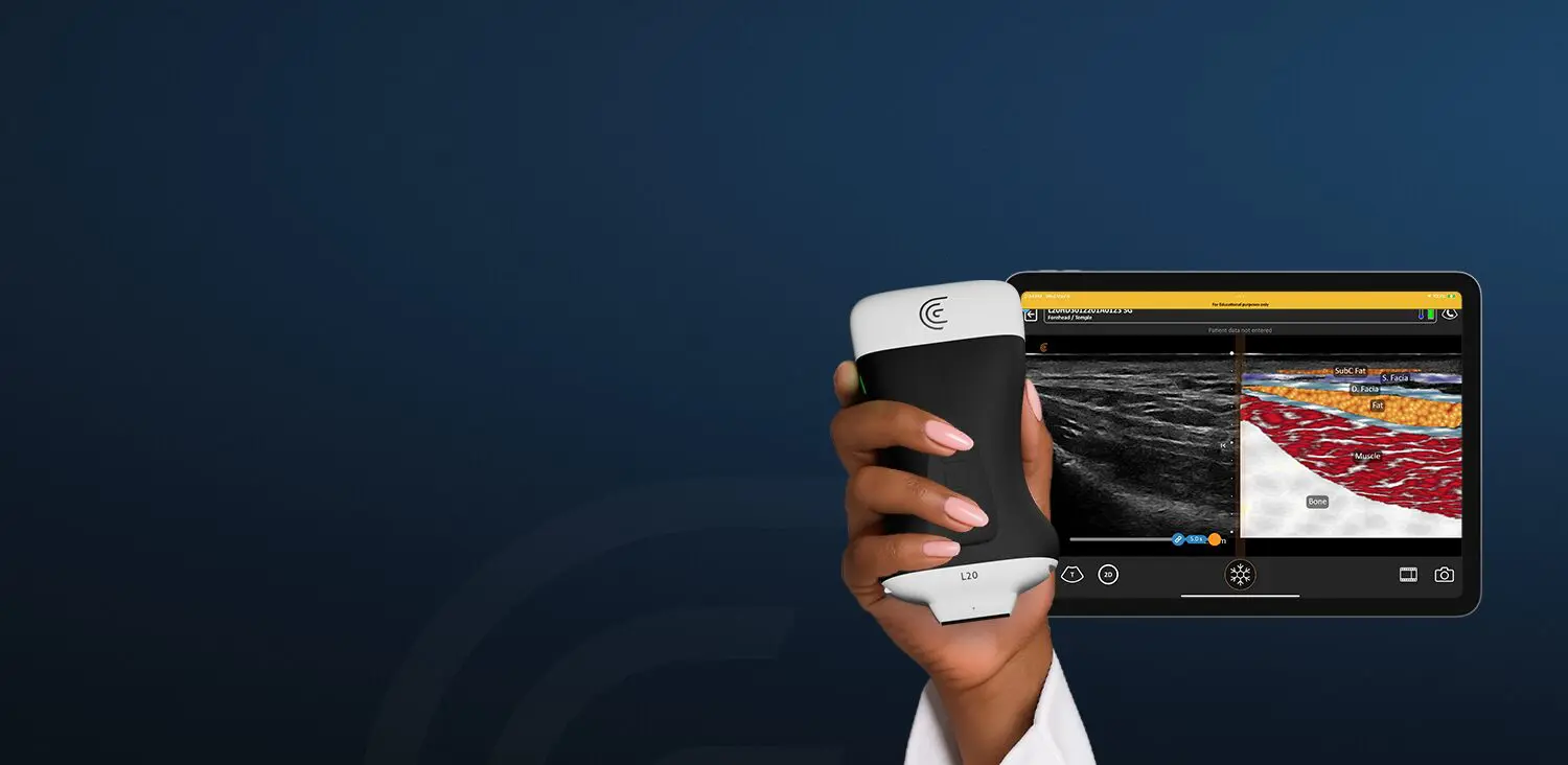
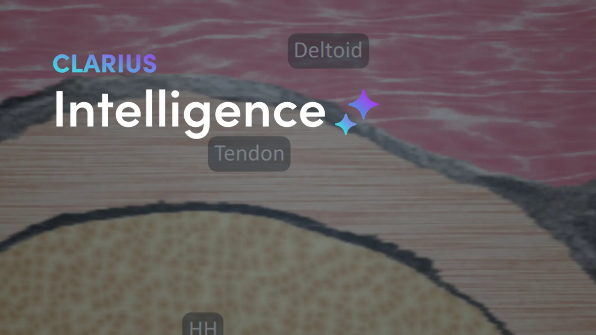
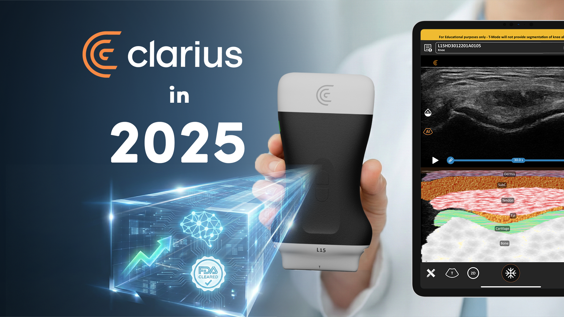
Referring to Clarius T-Mode as a « game-changer » in helping students with image interpretation, Dr. Voight was enthusiastic about using Clarius AI-powered tools to shorten the ultrasound learning curve.
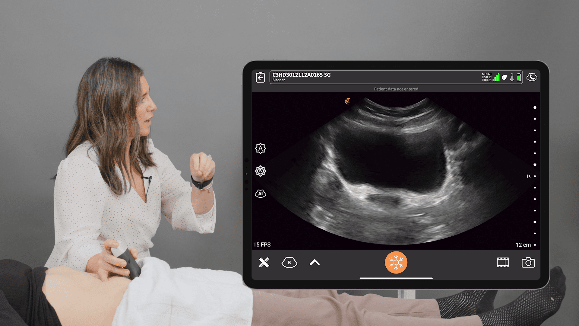
As demonstrated by webinar host Shelly Gunther, T-Mode provides a real-time, labeled graphical overlay of anatomical structures on the ultrasound image. This feature makes the initial learning process much less intimidating for students. As one resident explained in a video clip filmed during training at Belmont, T-Mode technology allows students to « learn it a lot faster » and get immediate confirmation of the structures they are viewing, decreasing their reliance on professors for verification.
Watch this video to see how T-Mode works.
Elevating Clinical Practice
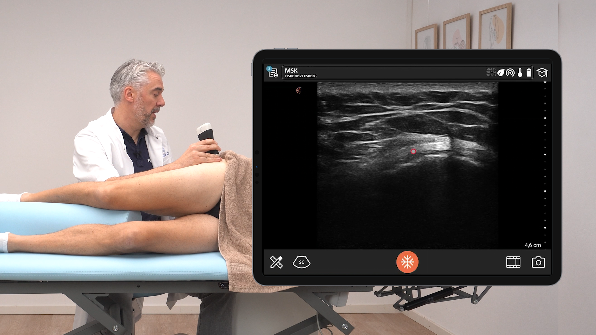
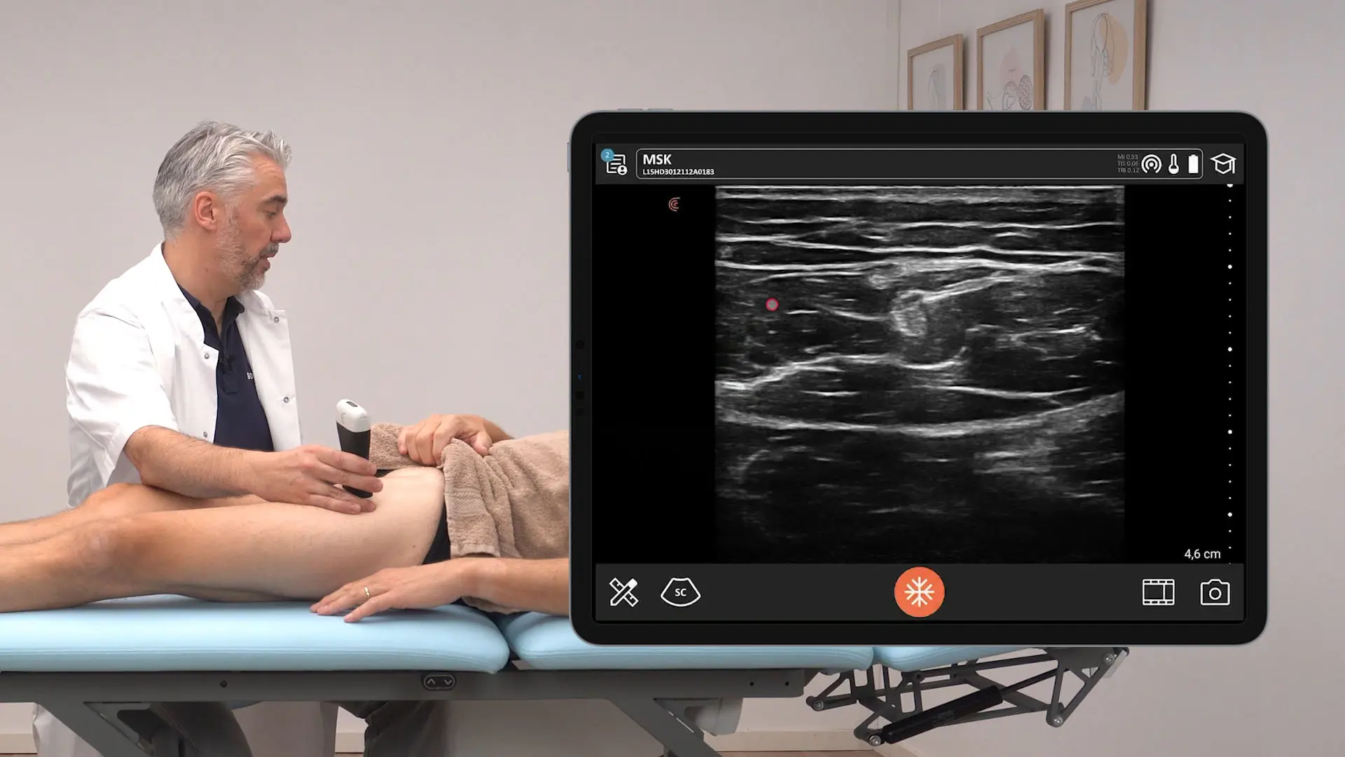
The integration of ultrasound training is not just an academic exercise; it has a direct and powerful impact on clinical practice. Dr. Wolfe, who also maintains a clinical practice, shared how ultrasound provides a more holistic view for clinicians and enhances patient care. It helps to increase patient buy-in and compliance by visually demonstrating the severity of an injury, treatment progress, or the accuracy of needle placement during procedures like dry needling.
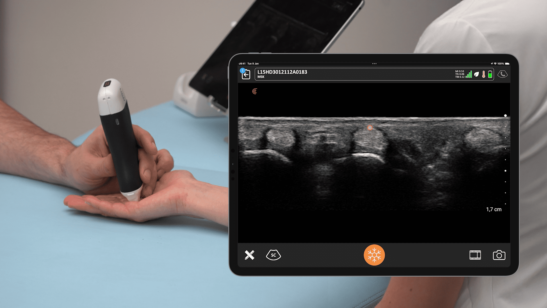
Watch this 2-minute video to see Dr. James Escaloni demonstrate his technique for accurate dry needling of the quadriceps tendon insertion onto the patella using the Clarius L15 HD3 high-frequency linear scanner.
Highlights from the Webinar Q&A
Question: What are the biggest challenges with integrating ultrasound into MSK education?
Answer: A pre-webinar poll revealed that the biggest challenges are limited training opportunities and a lack of access to ultrasound equipment. Dr. Michael Voight added that a survey of physical therapy programs found that a lack of qualified instructors and equipment costs were the primary barriers.
Question: Is there evidence to support the use of ultrasound in a physiotherapy clinic?
Answer: Yes, there is a large amount of easily accessible literature and research on scanning, sonography, and normative data for tissue pathologies.
Question: How do Dr. Voight and Dr. Wolfe use ultrasound in clinical practice?
Answer: Dr. Michael Voight’s clinical practice focuses on hip preservation, and he uses ultrasound daily in his hip clinic, noting that it has reduced the reliance on other imaging tools like MRIs.
Dr. Chris Wolfe primarily uses ultrasound for the hip (anterior, lateral, and posterior) for both assessment and treatment. For the spine, he uses it mainly for identifying the multifidus muscle in the cervical and lumbar spine. He also teaches safety measures during dry needling courses.
Question: Which Clarius model is best for hip ultrasound exams?
Answer: The Clarius C3 curvilinear scanner is recommended for imaging the hip because it provides greater depth. However, Clarius L7 linear scanner can also provide good images, depending on the patient’s size and the desired field of view.
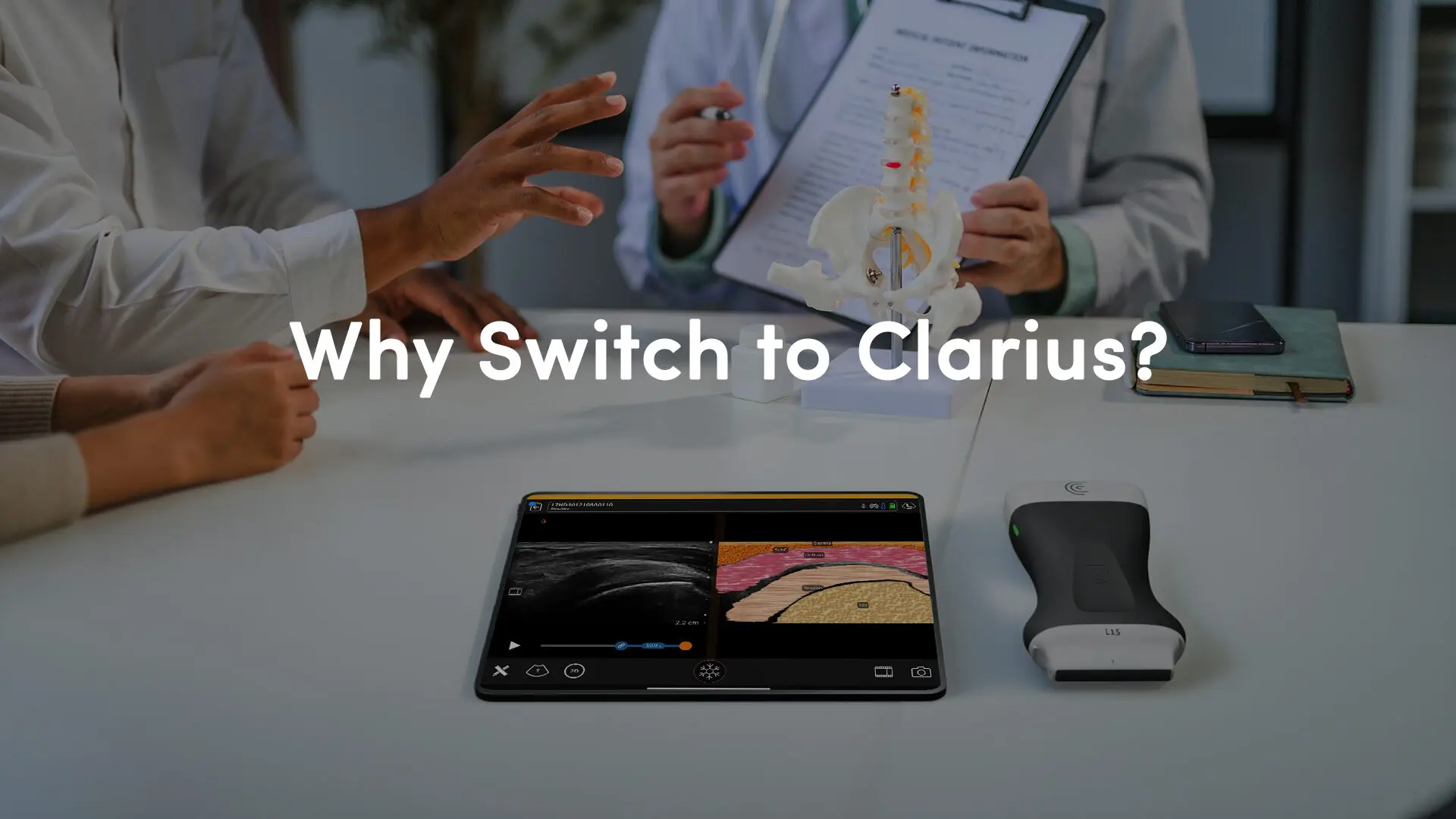
Curious About Clarius MSK Ultrasound for Your Institution or Practice?
If we’ve piqued your interest in discovering whether Clarius ultrasound is right for your practice or teaching institution, we invite you to book a virtual demonstration. We’ll be delighted to show you why Clarius ultrasound is the leading choice of handheld ultrasound for discerning clinicians.

