


As a physiotherapist, do you wish you could show your patients what’s happening inside their bodies when you ask them to engage muscles they can’t see or feel? For many pelvic health physiotherapists, this has been a significant barrier to effective treatment. The good news is that ultrasound technology is now more readily available for physiotherapists to make the « invisible » visible. Affordable and easy-to-use, intelligent handheld ultrasound scanners are a game-changer for pelvic health physiotherapy, enhancing both assessment and patient education.
We recently hosted a webinar with pelvic health physiotherapist Adrienne Sim where she demonstrated the versatility and accessibility of ultrasound for clinical practice. Since ultrasound has become readily available in the clinics where she worked, « it has become a core part of how I practice,” she says. The transabdominal approach is non-invasive, making it a comfortable and accessible option for a wide range of patients, especially when internal exams are not appropriate or desired due to trauma, discomfort, or medical contraindications. As Adrienne explains, using ultrasound « is especially valuable when internal exams are not appropriate or desired by the client ».
Watch the free one-hour webinar at your convenience: POCUS for Pelvic Health: Enhancing Assessment & Treatment with Transabdominal Real-Time Ultrasound. Read on for highlights and a demonstration video.
Clinical Highlights: The Benefits of Ultrasound for Pelvic Health Physiotherapy
- Improved Patient Education and Engagement: Seeing their own anatomy in motion on a screen helps patients connect with their body, understand instructions, and feel empowered in their treatment. Adrienne says, « The visual feedback helps to improve motor learning and helps clients engage more effectively and potentially progress more quickly ».
- Objective Insights without Internal Exams: For clinicians who do not perform internal exams, transabdominal ultrasound offers a valuable tool to visualize and assess pelvic floor and core strategies, providing objective insights that help guide care.
- Real-time Biofeedback: Ultrasound serves as a valid form of biofeedback to guide pelvic floor muscle training, a first-line treatment for many pelvic floor conditions. As Adrienne states, « We can also back up what we’re observing through palpation or functional testing with real-time visual imaging, which adds clarity and credibility to our assessments ».
- Versatile and Adaptable Assessments: Assessments are possible in a range of different positions, including lying down, sitting, standing, and even on hands and knees, making them functionally relevant and adaptable to different patient needs and activities.
- Assessment of Pelvic Floor Muscle Activity and Coordination: Ultrasound allows clinicians to observe the elevation of the bladder base during a pelvic floor muscle contraction. It can also reveal dysfunctional recruitment patterns, such as a lowering of the bladder base, and provides insight into the quality of relaxation.
- Core Muscle Assessment: It is one of the only clinical methods available to visualize the morphology and behavior of the transverse abdominis in action. As Adrienne explains, this feedback is « especially validating for clients who aren’t sure if their core is activating correctly, and it helps to bridge the gap between sensation and function for our patients ».
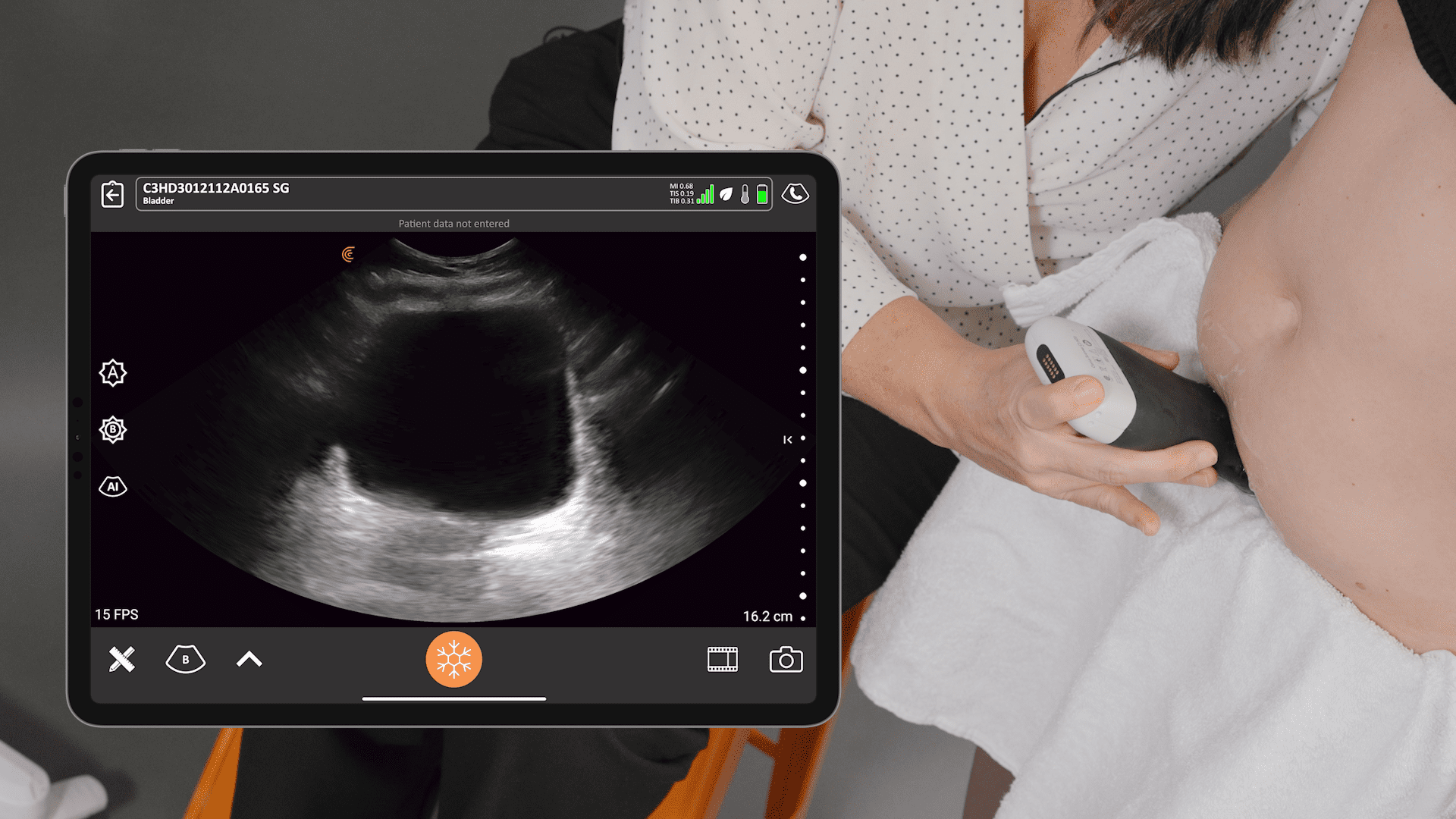
- Bladder Function Evaluation: Ultrasound can be used to measure bladder volume and post-void residuals (PVR). For patients with urinary urgency, visualizing bladder volume in real-time can help them better conceptualize their bladder sensations and retrain their perception of urgency.
[VIDEO] Ultrasound Demonstration: Functional Pelvic Floor Assessment – Standing
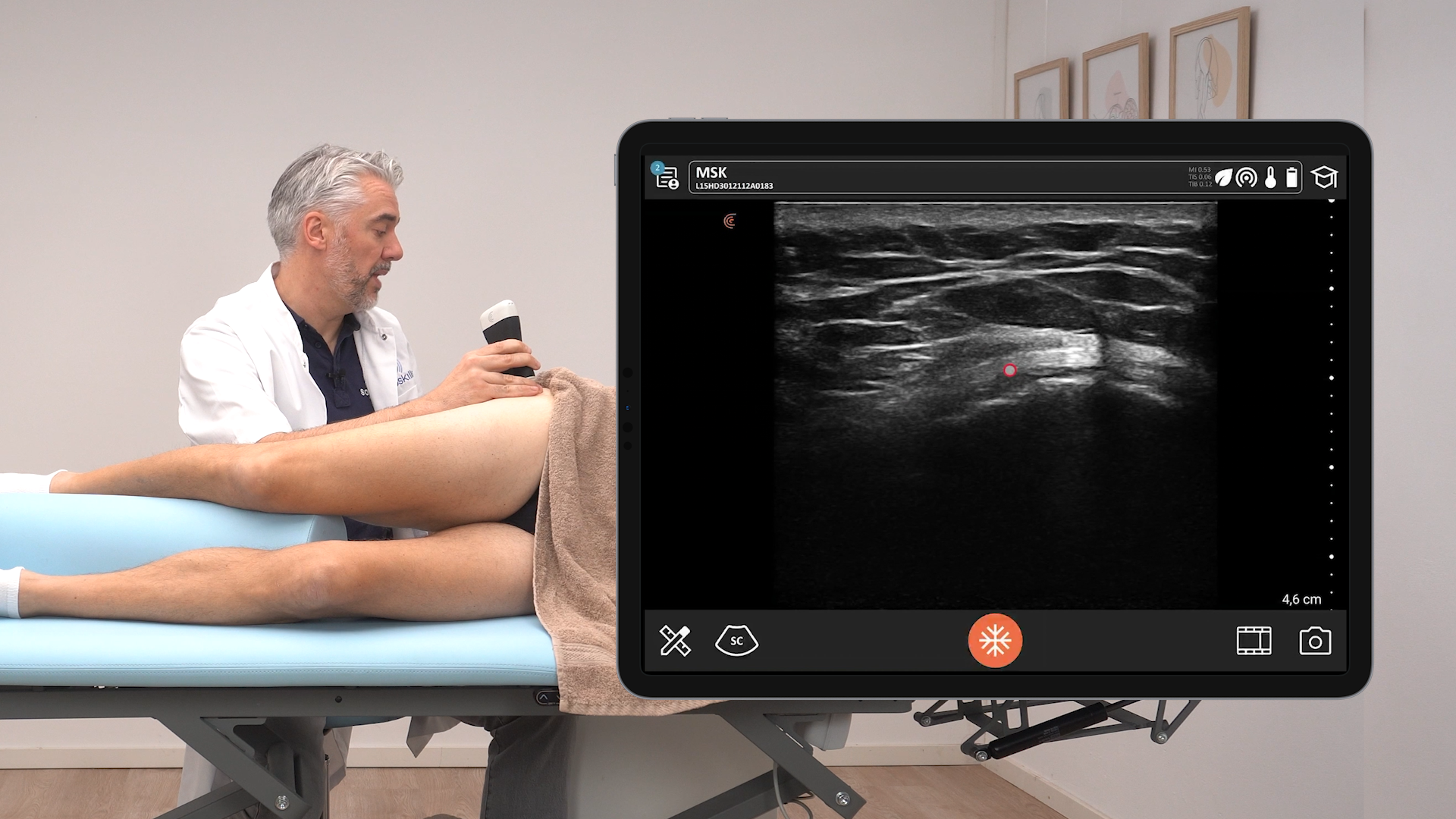
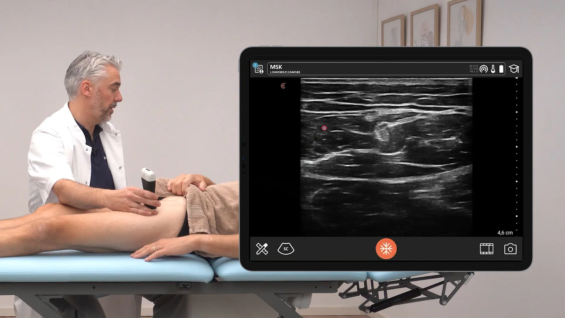
In this 2-minute video, Adrienne demonstrates the ultrasound appearance of pelvic floor contraction, endurance hold, and coordinated contractions with her client in a standing position using the Clarius C3 HD3.
[Video] Ultrasound Demonstration: Functional Pelvic Floor Assessment – Supine
Watch Adrienne scanning her patient’s abdomen with the Clarius C3 HD3 while she performs pelvic floor contractions, endurance holds, and coordinated contractions in a supine position. Visit Clarius Classroom for more videos.
Q&A
Q: Is ultrasound effective for female pelvic health, and are there any side effects, especially for pregnant women?
A: Adrienne Sim uses ultrasound « very frequently for pelvic floor assessments ». It is a valuable tool for a varied range of conditions including incontinence, prolapse, pelvic pain, and pre-and post-natal rehab. She did not mention any side effects.
Q: How do you instruct a patient to engage their pelvic floor muscles during a contraction test?
A: Adrienne suggests that « one of the best cues that’s been shown to translate onto ultrasound is more of a puborectalis cue ». She elaborates that this involves the idea of « squeezing the anus or holding wind » to create the upward motion that is imaged on the transverse view.
Q: How accurate is this technology for morbidly obese patients?
A: Adrienne Sim explains that it « can be more difficult to image with a greater abdominal circumference ». However, the curvilinear probe offers greater depth penetration and can be used for clients with a larger abdominal circumference or higher body mass index.
Q: When assessing pelvic floor muscles, are there any standardized measurements for monitoring progress, or is it a subjective assessment?
A: While some research analyzes the amount of bladder base displacement, Adrienne notes that in clinical practice, she doesn’t typically do that. Instead, she focuses on the « quality of the contractions, » observing the « symmetry of the contraction, the symmetry of the relaxation, the completion or lack of completion of the relaxation ». She also assesses the ability to perform a sustained endurance hold or quicker, coordinated contractions, which is important for voluntary control during tasks like coughing or sneezing.
Q: Is there a standard measure or ratio for the transverse abdominis?
A: According to the live demo, the transverse abdominis should be about half the girth of the internal oblique. It is a postural muscle, so it will always have a lesser girth than the internal and external obliques.
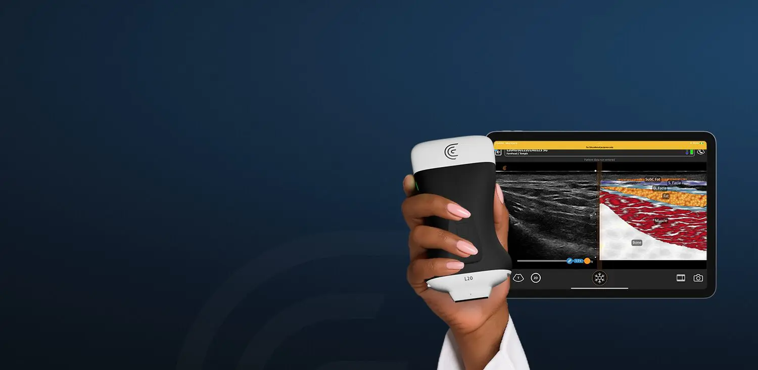
The Clarius Advantage for Physiotherapy
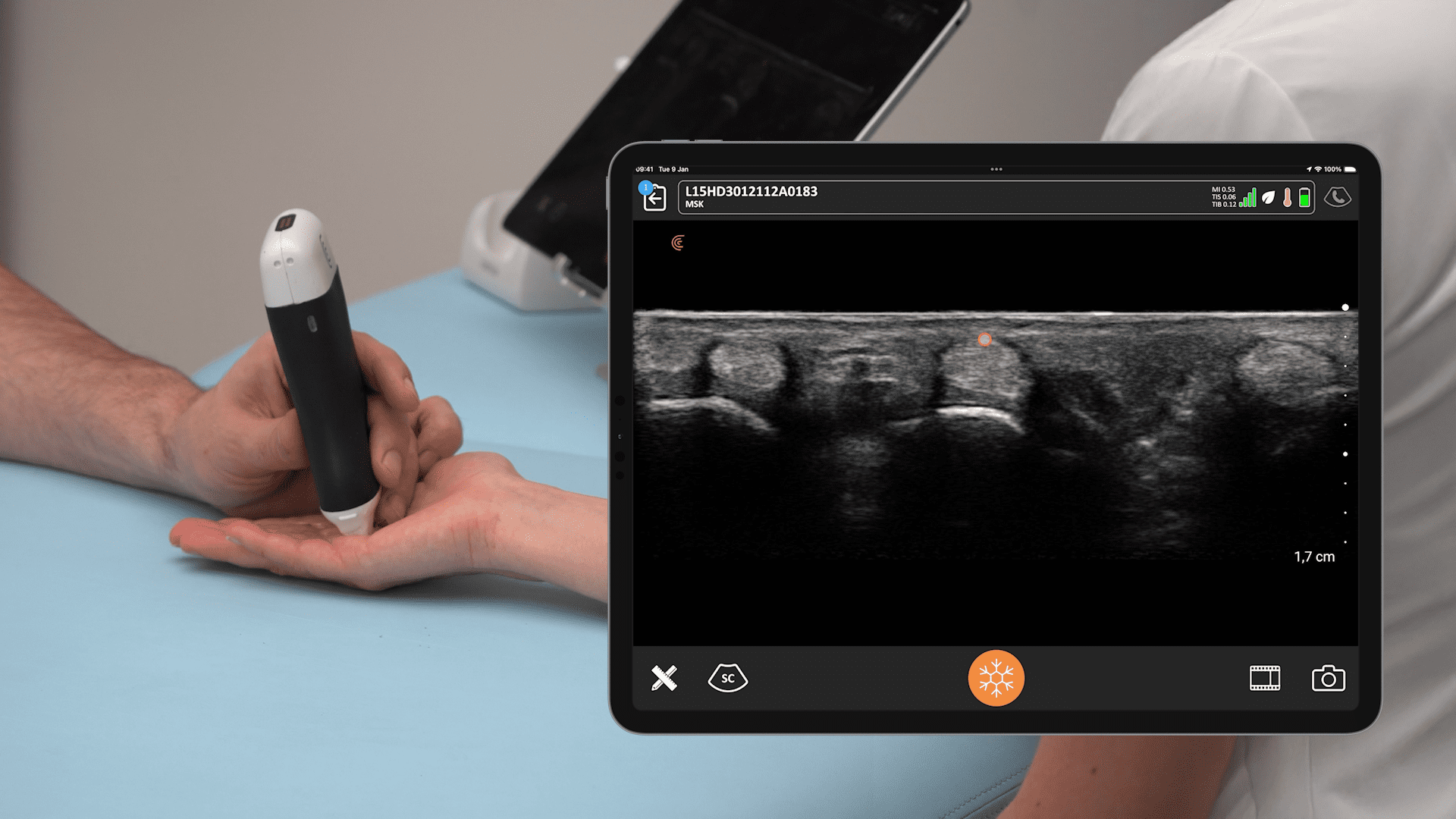
The Clarius HD3 is the third generation of portable ultrasound scanners that can be used for physiotherapy. Cost-effective, compact, and easy to use, Clarius scanners are ideal for clinics where space is limited. Their cordless design allows for a smoother workflow and more comfortable interaction with clients compared to a traditional cart-based or corded system.
The Clarius ecosystem includes several features that enhance its usability and effectiveness:
- Free App: A free app for iOS or Android devices allows for unlimited users.
- Clarius Cloud: This feature is easily used to capture and manage unlimited exams, making it a great tool for saving images and following a patient with serial studies.
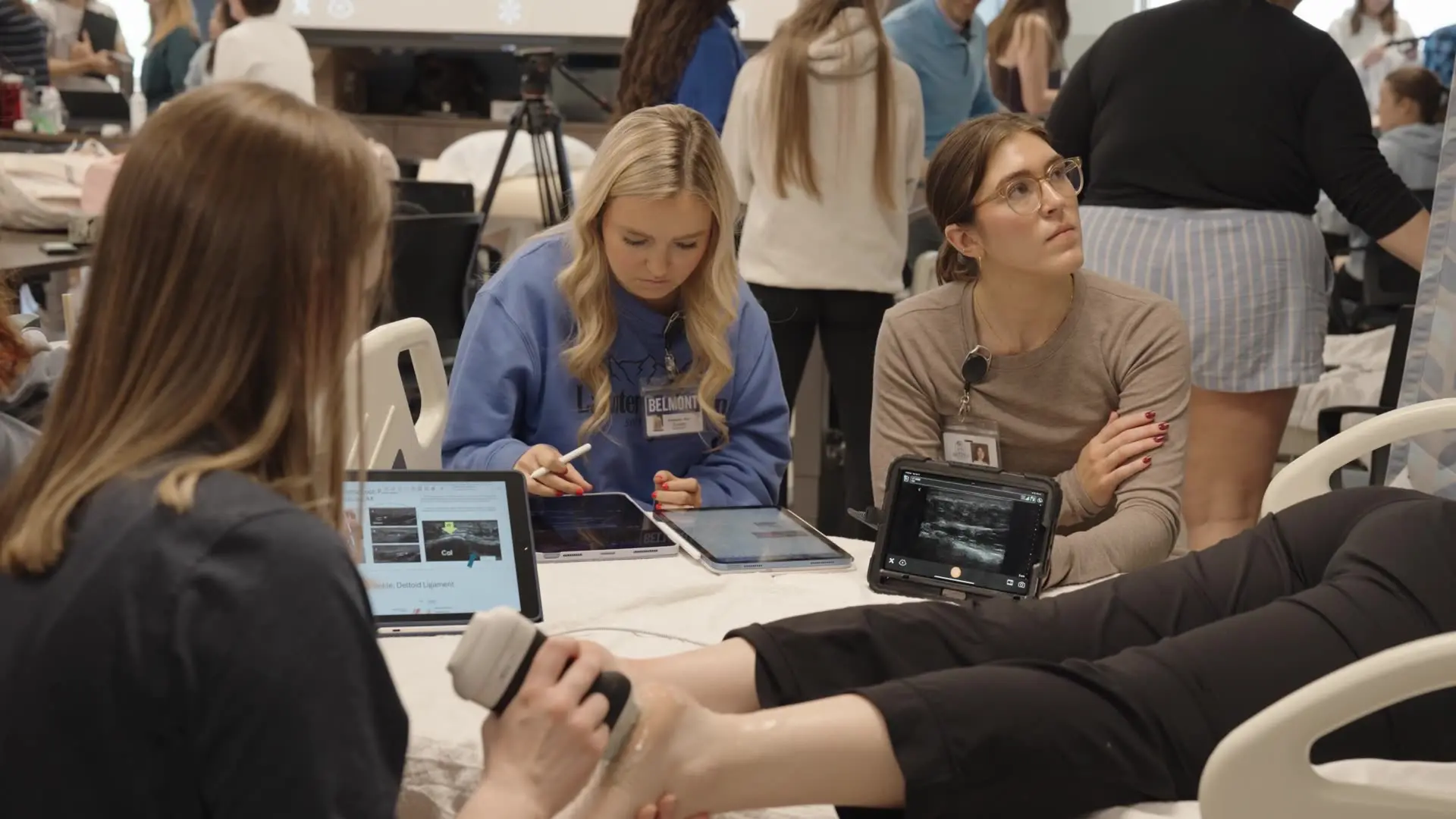
- Clarius Classroom: The membership includes in-app videos and onboarding with a clinician to help users build their skills.
- Clarius Live: This provides one-click telemedicine, allowing users to share live scanning with a colleague for a second opinion.
- Advanced Features: The membership also includes advanced features such as dedicated presets, bladder AI, and voice controls.
- Wireless and Portable: The wireless design frees up space with a zero footprint and offers ultra-portability, allowing for free movement without wires.
To learn more about how easy and affordable it is to add Clarius HD3 to your practice, contact us today or request a personalized virtual ultrasound demo.

