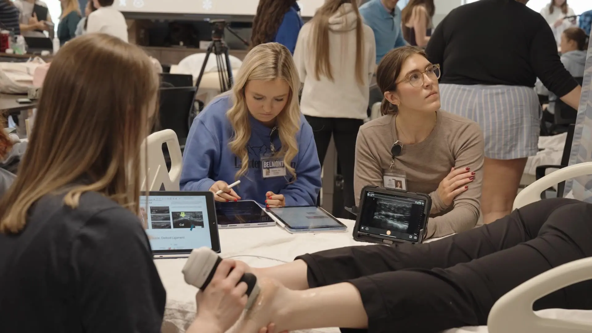
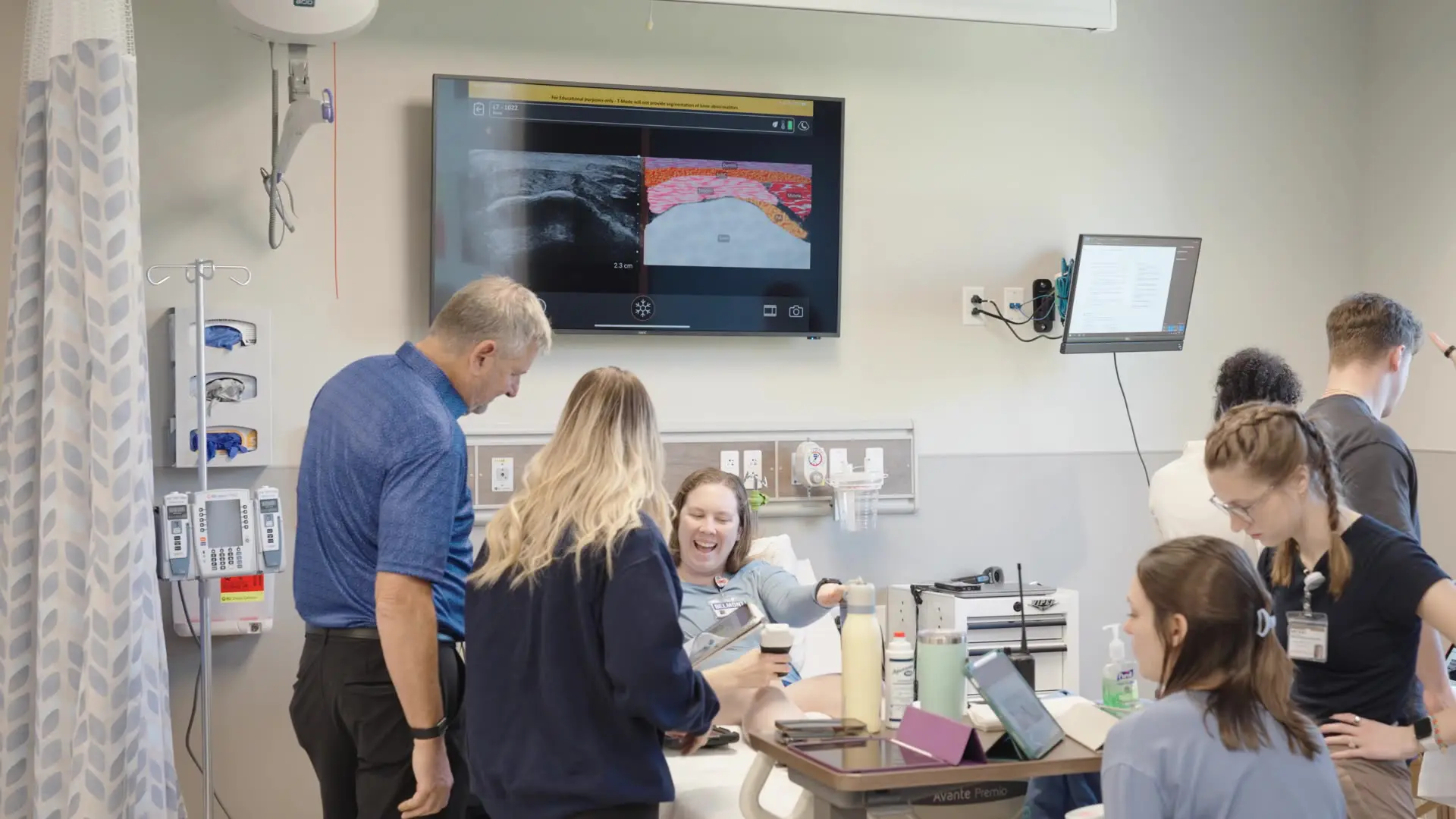
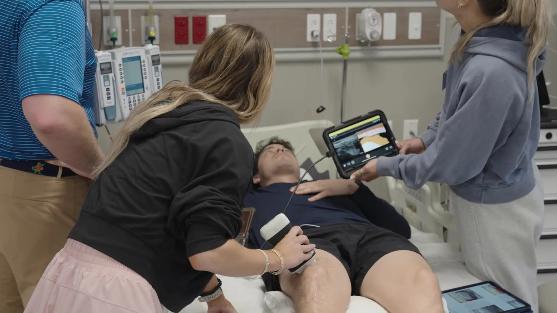
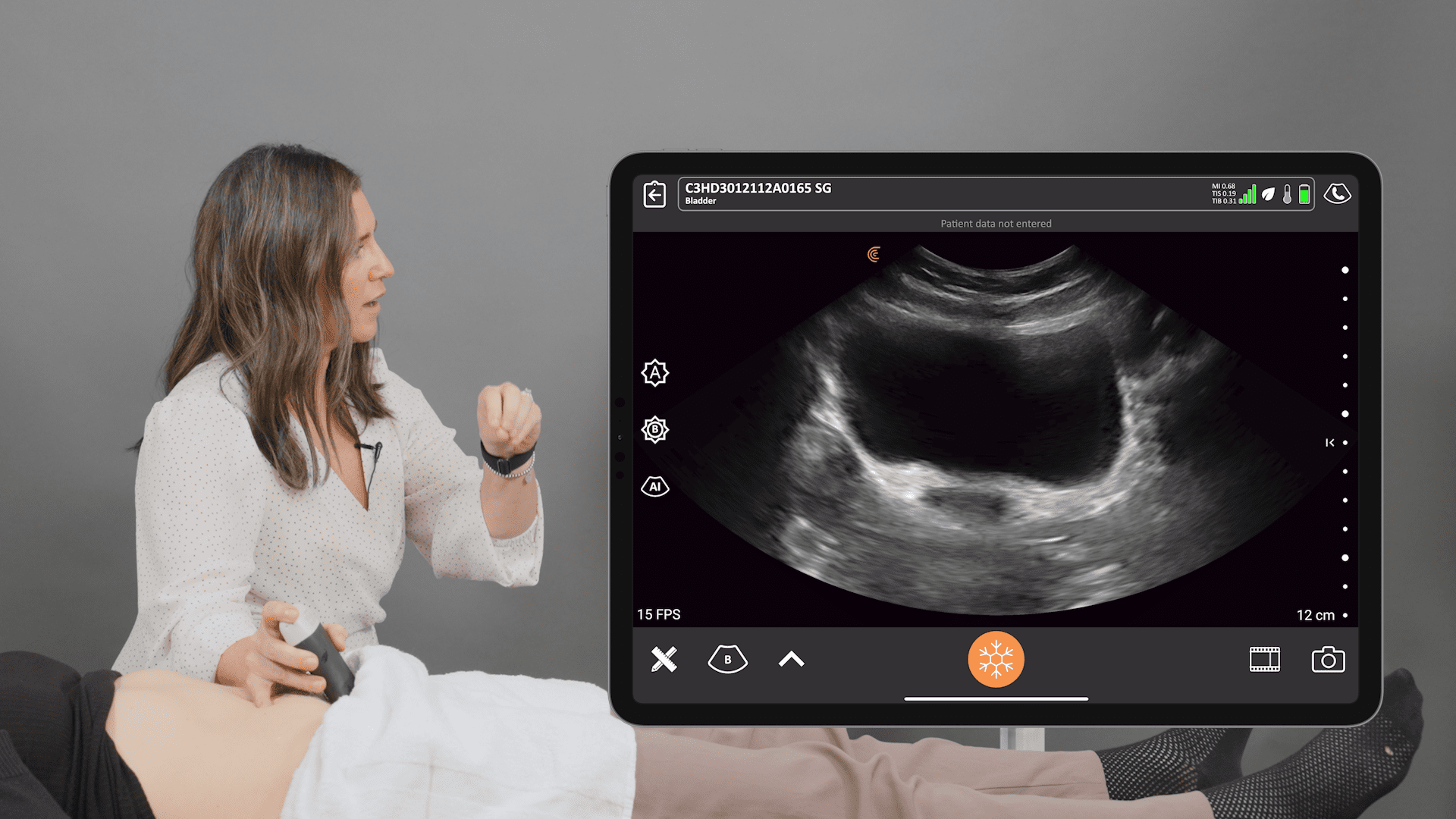
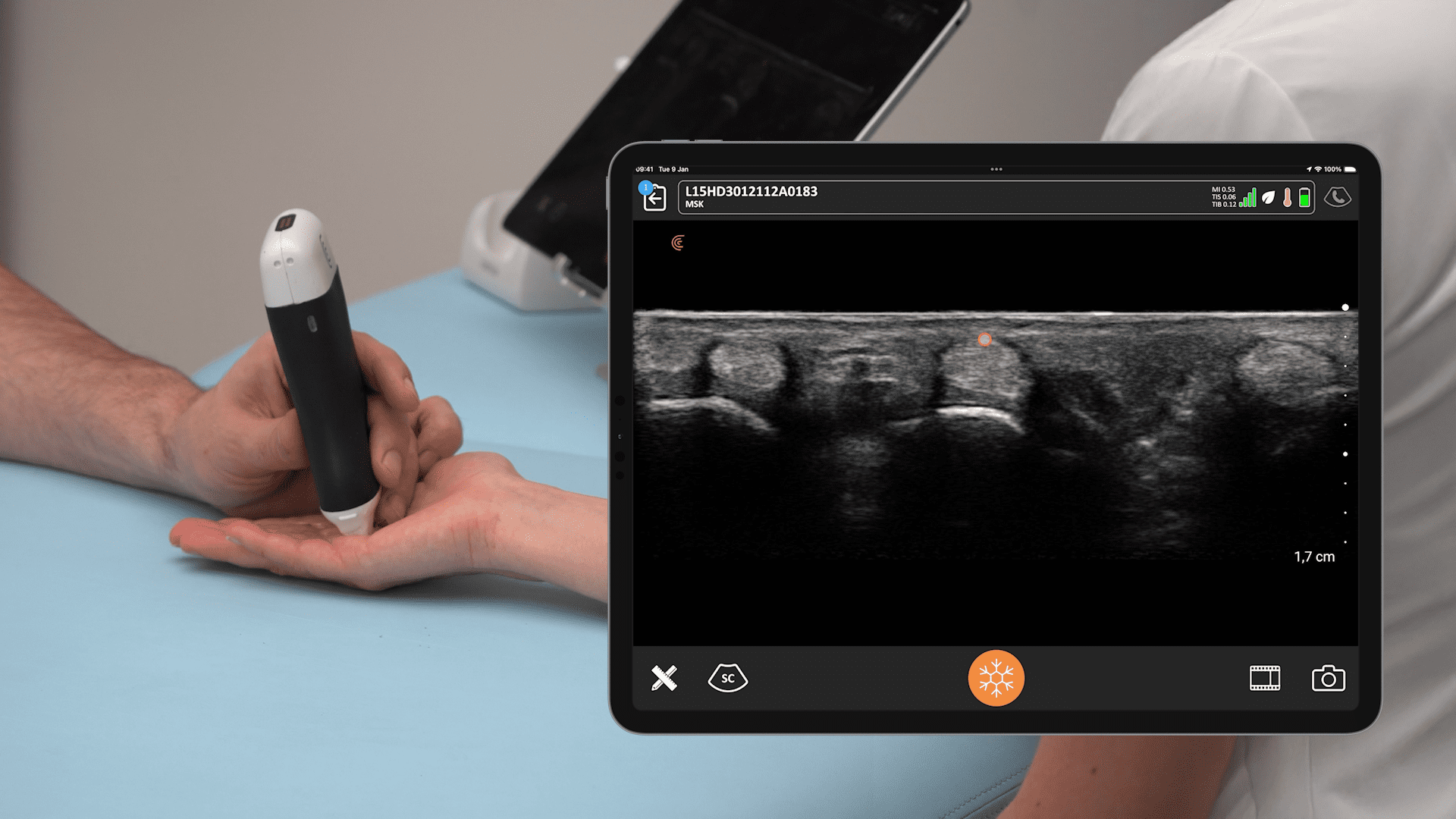
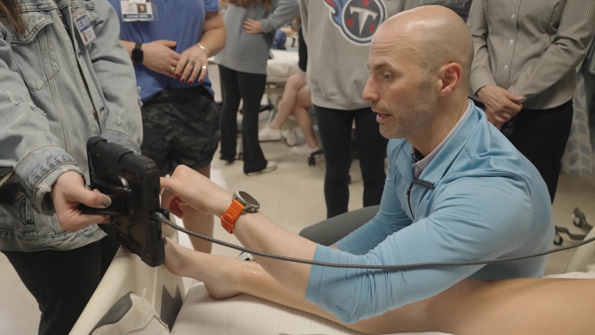
There is increasing evidence in the literature that states ultrasound guidance is becoming the standard of care for regional anesthesia, and contributes to better, safer outcomes for patients. Regional anesthesiologist Dr. Greg Hickman from the Andrews Institute Ambulatory Surgery Center in Florida has twenty years of first-hand experience using ultrasound for better patient outcomes. He joined us for another instructional webinar, « Ultrasound-Guided Brachial Plexus Blocks: Techniques from the Expert! ». We invite you to watch the full webinar to improve your handheld ultrasound skills and perfect guided blocks for upper extremity surgery or read on for highlights from Dr. Hickman’s presentation.
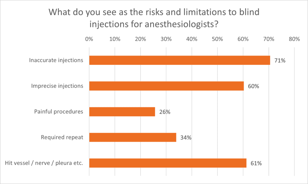
Early in the webinar, we conducted a poll asking viewers their thoughts regarding performing blocks blindly, and it’s evident that inaccurate, imprecise injections as well as complications are the top concerns.
Dr. Hickman discusses and demonstrates 3 upper extremity blocks for surgeries below the shoulder, using dual guidance with ultrasound and a nerve stimulator.
The Supraclavicular Brachial Plexus Block
Dr. Hickman calls this block the “Spinal of the Arm”. It’s his “go-to” block at the Andrews Institute as it proves rapid and highly effective analgesia for all upper extremity surgeries. This block happens at the level of the divisions of the trunks
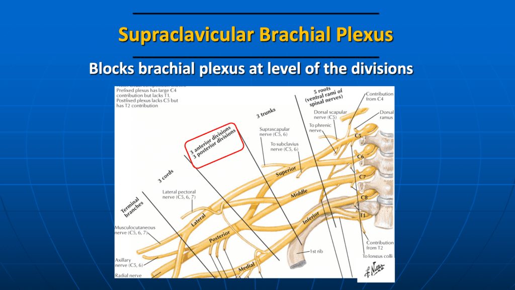
Anatomical landmarks are the clavicle and the sternocleidomastoid. On the ultrasound, landmarks are the subclavian artery and the 1st rib.
“Find the group of grapes just posterolateral to the subclavian artery, and that’s our go-to”.
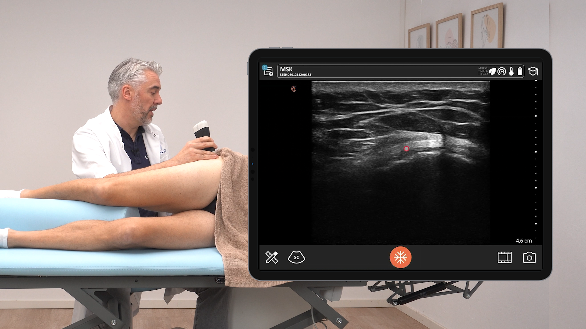
Watch this Clarius Classroom video to see how Dr. positions his patient and the ultrasound scanner, locates the supraclavicular brachial plexus, and performs the block with a total of 30 cc’s of analgesia injected to surround the trunks.
The Infraclavicular Brachial Plexus Block
This block is at the level of the lateral, medial and posterior cords, and like the supraclavicular block, is predictable, has fast onset, and is effective for the whole arm and hand. During the webinar, Dr. Hickman explains why this is his preferred block when using a continuous catheter.
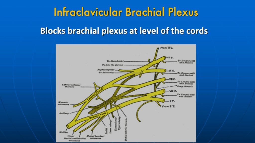
With the scanner in long axis, and placed in the deltopectoral groove, anatomical landmarks include the inferior aspect of the clavicle and medial to the coracoid process. Locate the axillary artery to identify the cords of the plexus.
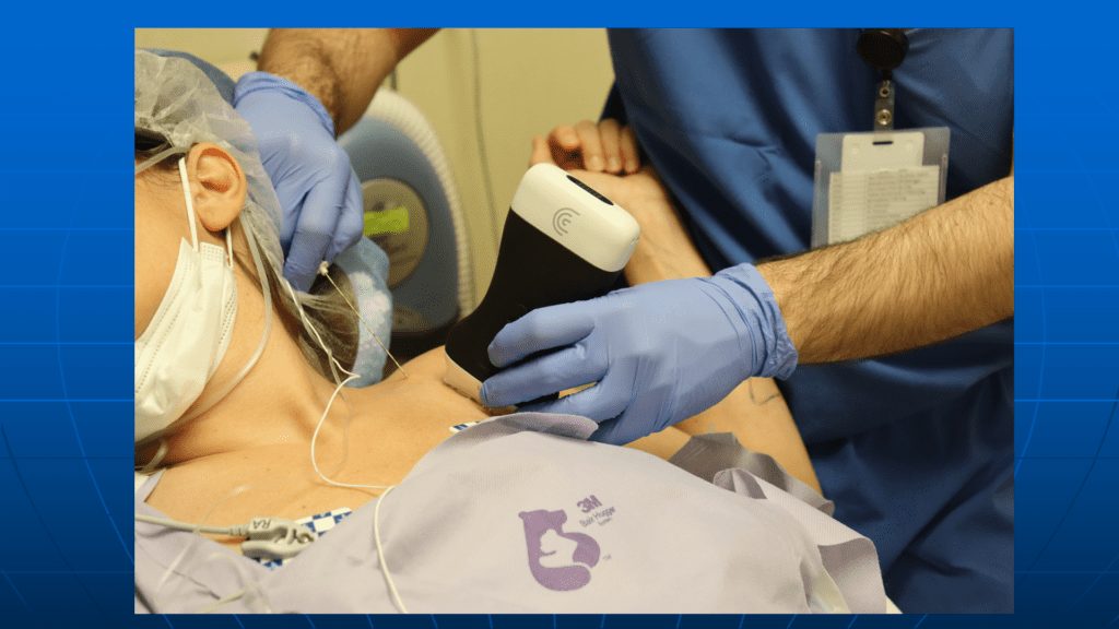
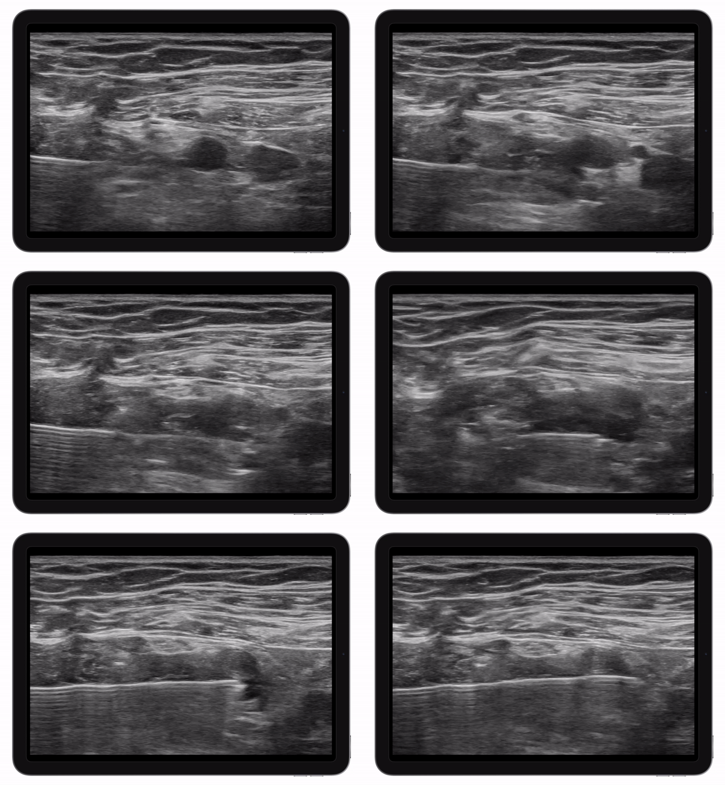
Dr. Hickman uses a retroclavicular approach for this injection because there’s no special patient positioning, and he can visualize his needle in-plane, at an optimal angle, as it appears from beneath the clavicle.
Slight needle manipulation under ultrasound guidance ensures anesthetic deposit around each cord.
Watch the video:
The Axillary Brachial Plexus Block
In cases where a supra- or infraclavicular block is technically challenging, the axillary block can be very effective and easier to perform because of the superficial nature of the plexus. The radial, ulnar, median, and musculocutaneous nerves can be easily identified with ultrasound, and the needle is highly visible with the angle of approach Dr. Hickman uses, making the injection accurate and effective.
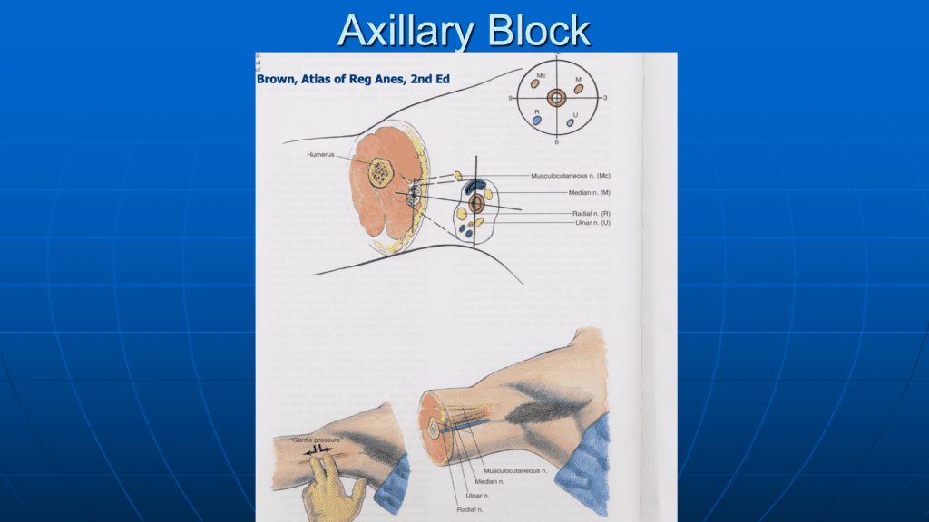
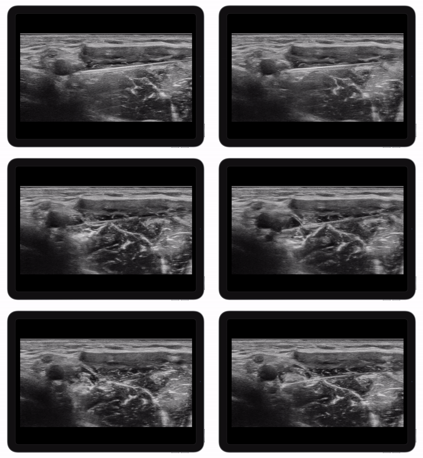
The key ultrasound landmark for this block is the axillary artery, imaged in short axis. The radial, ulnar and median nerves lie in close proximity to this prominent vessel, and the musculocutaneous nerve can be seen close by in the intermuscular septum between the biceps brachii and brachialis muscles.
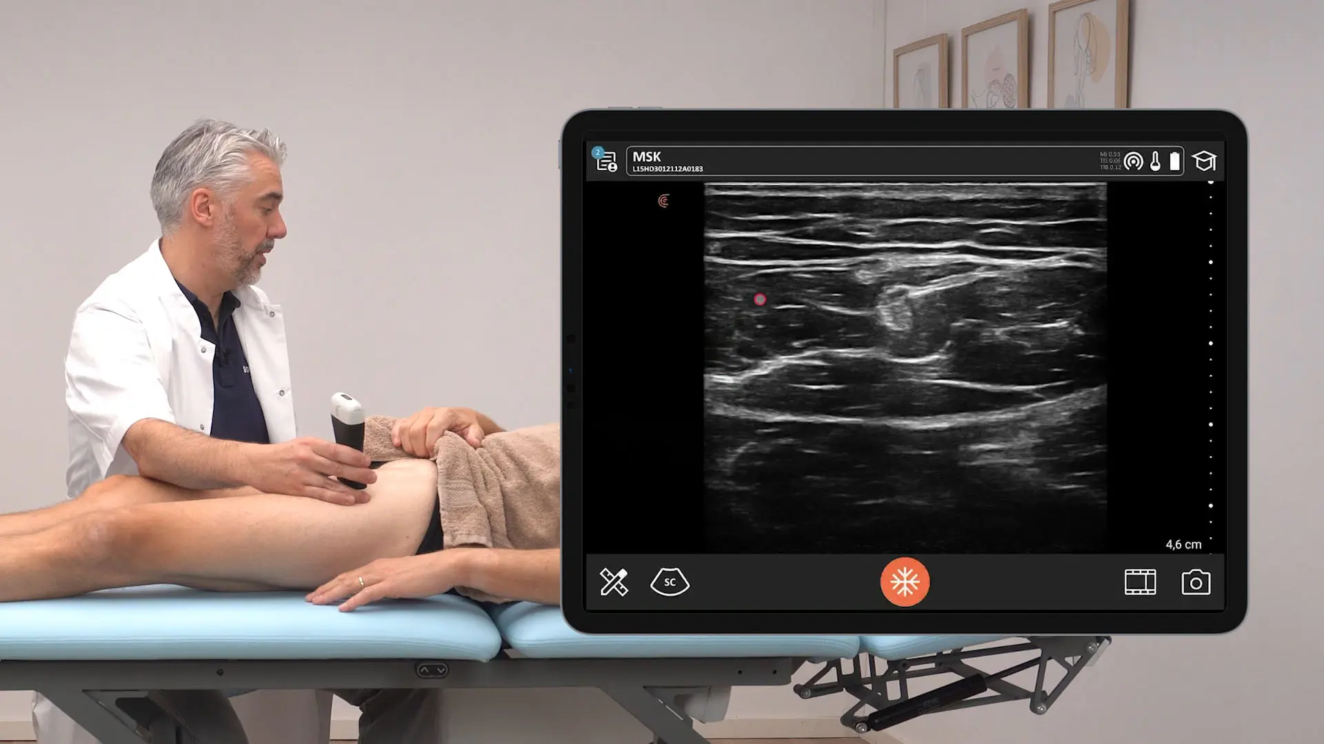
Watch this live video to see the median and ulnar part of this block.
Dr. Hickman has been using ultrasound to guide his blocks for 15 years now.
I didn’t realize the true impact of how much it was going to change and how many more people would get into regional anesthesia because so many people can feel more comfortable with this than trying to do those blind techniques,” he says.
For more detailed instruction and full brachial plexus block demonstrations watch the full webinar, « Ultrasound-Guided Brachial Plexus Blocks: Techniques from the Expert! ». Visit the Clarius Classroom for these and other regional anesthesia block procedures.
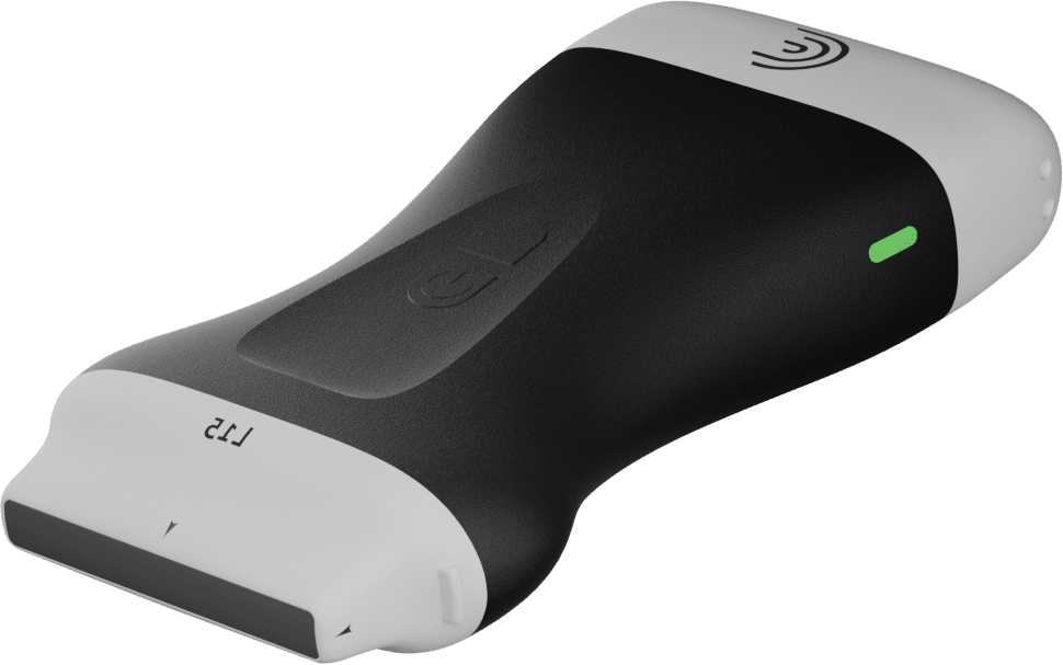
Dr. Hickman is co-founder of the popular ultrasound-guided regional anesthesia education website Blockjocks.com, which teaches doctors, nurses, and patients around the world. He uses the Clarius L15 HD3 at his practice.
About Clarius for Regional Anesthesia
The miniaturization and innovation of handheld ultrasound mean high-definition imaging is now easy and affordable, offering savings of up to 80% on the cost of a traditional ultrasound system. Clarius offers three wireless handheld ultrasound scanners that are suitable for safe regional nerve blocks and post-operative follow-up. For more information, visit our anesthesia specialty page. Or contact us today to request a private virtual demonstration of the ultimate wireless ultrasound scanner.
