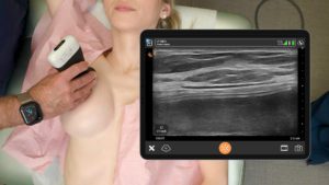
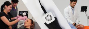
Dr. MJ Rowland-Warmann has been using the Clarius L20 at her practice since 2020. She is the founder and lead clinician at Smileworks Hub in Liverpool. Dr. Rowland-Warmann has a special interest in the management of dermal filler complications using ultrasound and is a huge advocate for safer aesthetic procedures and is working toward raising industry standards.
We recently co-hosted a lively webinar titled « Avoiding Filler Complications Using Ultrasound to Guide Safe Cheek, Temple, and Tear Trough Injections« . Watch the webinar or read on for a quick summary.
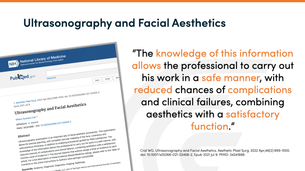
Ultrasound is the most important thing to come to aesthetics since the invention of dermal filler,” according to Dr. Rowland-Warmann. Literature citing research continues to support this assertion.
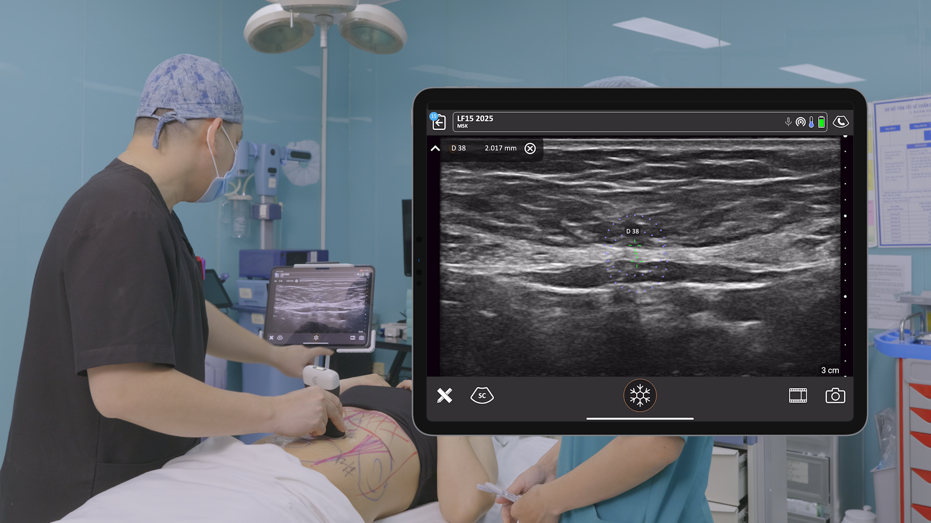
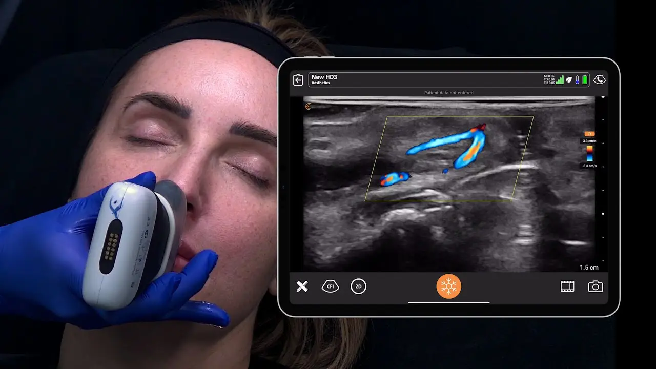
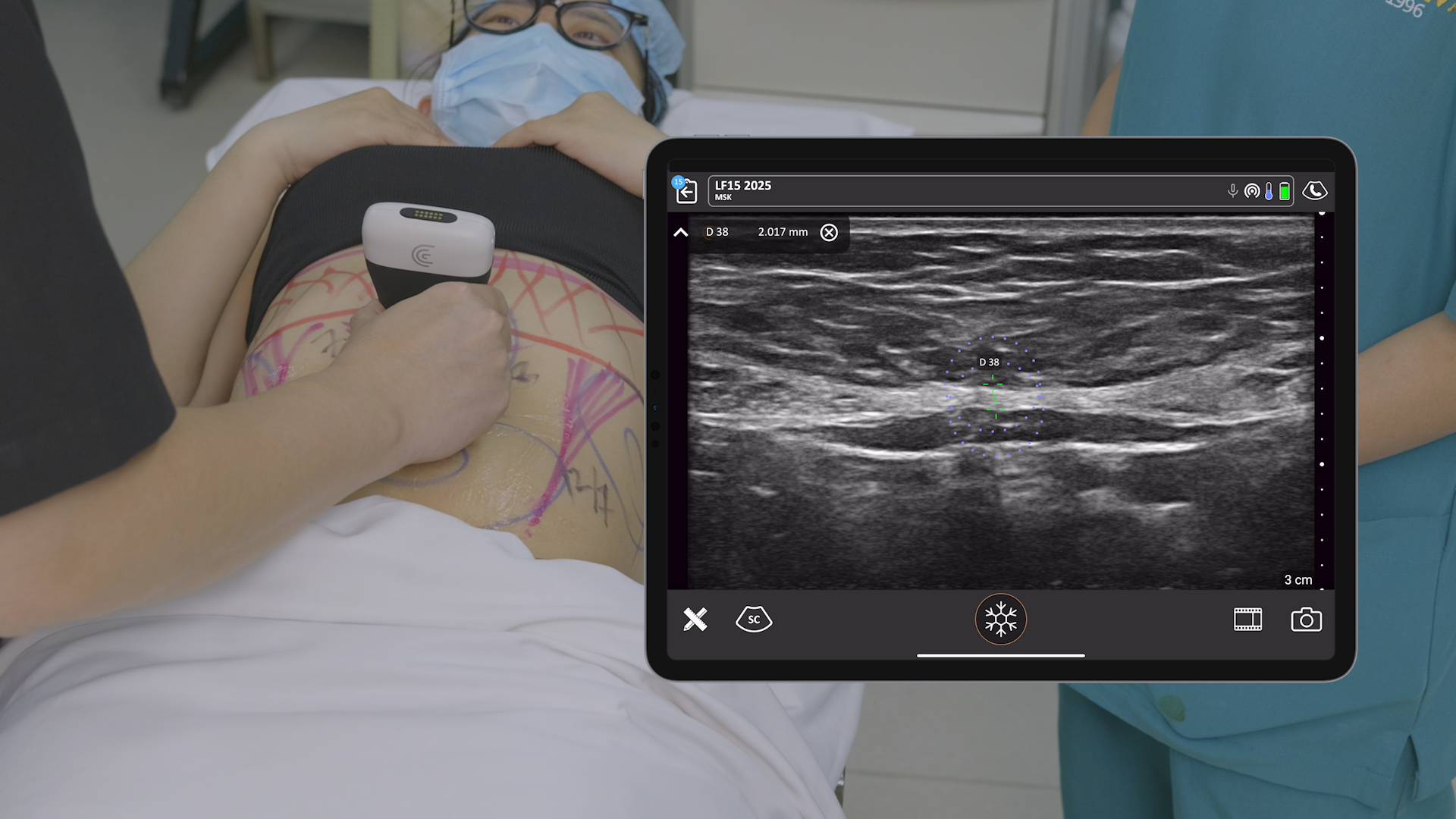
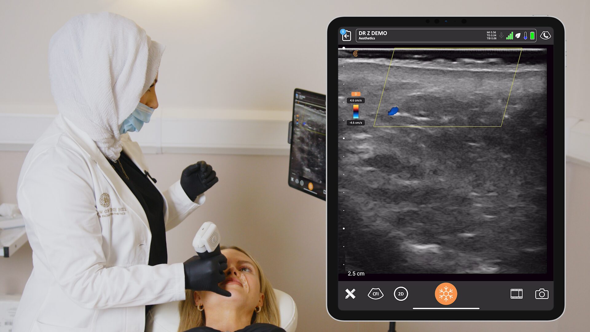
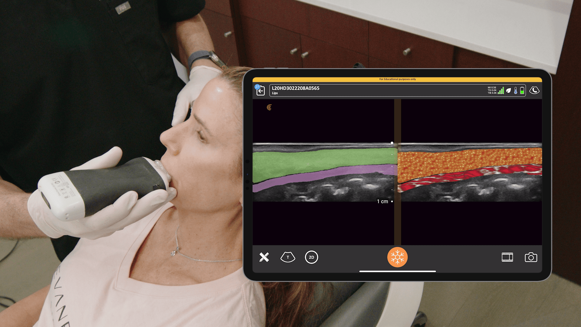
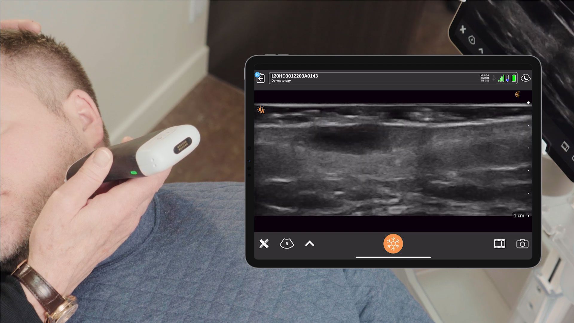
In the webinar, Dr. Rowland-Warmann describes the many benefits of using high-resolution ultrasound before, during and after filler procedures, and shares live video of ultrasound-guided procedures on two patients.
How Ultrasound Can Help Us
- Filler treatment – before, during and after
- Before – identify anatomy and perform vascular mapping, essential for treatment planning and safer injections
- During – also called guided therapy, ensuring precise and more predictable filler placement
- After – confirm filler placement and vascular integrity
- Diagnosis and management of complications
- Identify occlusions and treat them quickly and effectively
- Monitor filler over time – identify previously injected filler and manage your patient accordingly
- Diagnosing filler types – ultrasound appearance can vary with different fillers
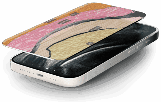
- Learning anatomy – identifying structures under the skin in real-time is invaluable when performing injections
Performing US Guided Temple Augmentation
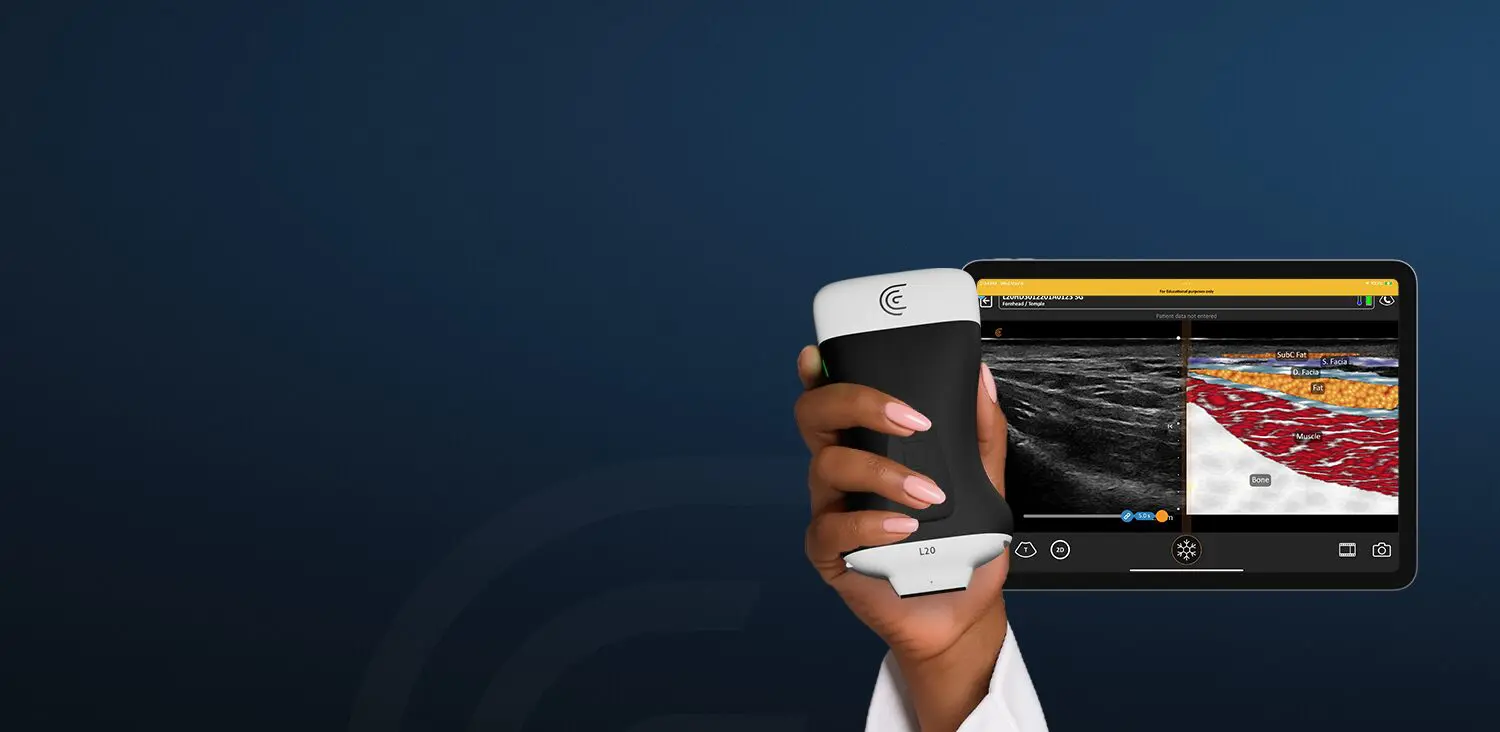
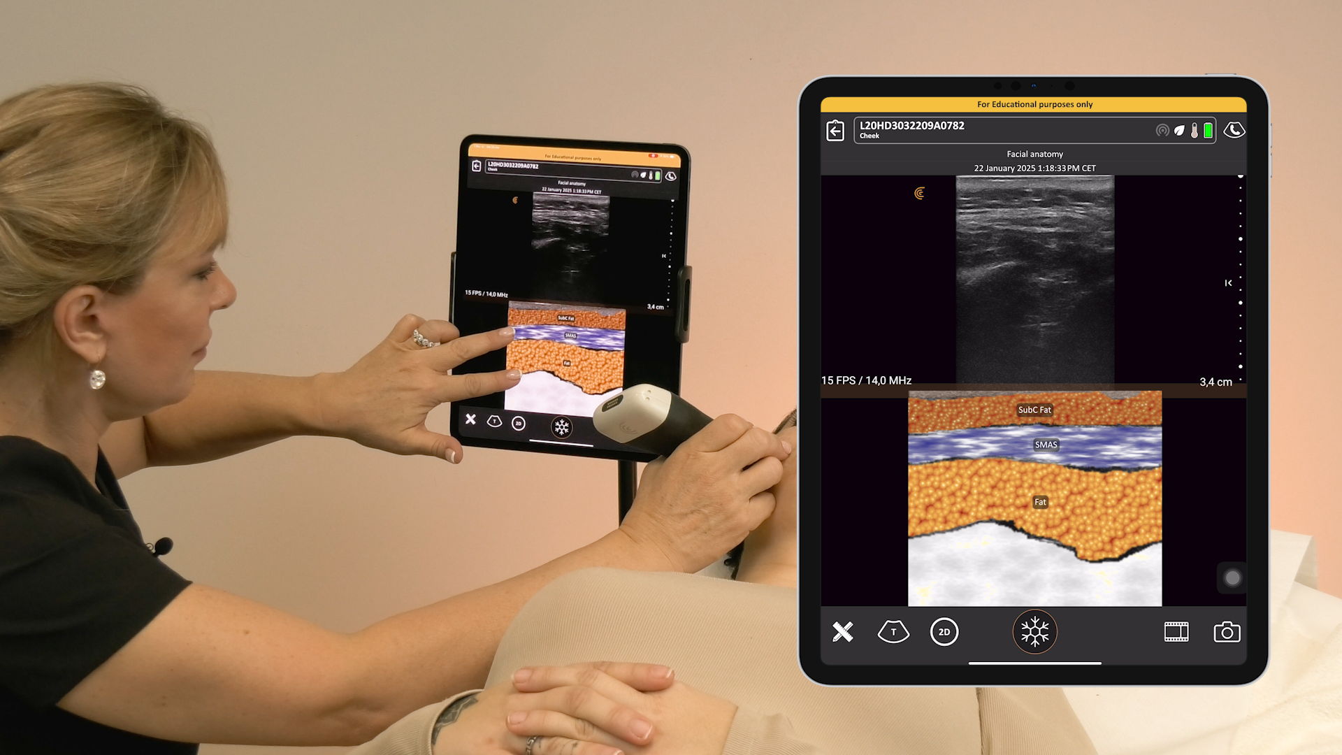
Although there are six different injection techniques for the temple, Dr. Rowland-Warmann prefers the interfacial technique. Watch the video as she describes the fascial layers of the temple, and the ultrasound appearance of the superficial and deep temporal fasciae. Color Doppler is a valuable tool to identify the superficial and deep temporal arteries in this area between these 2 layers is the deep fat layer, where the filler is placed with precision using ultrasound guidance.
Performing US Guided Cheek Injection
Treatment of the cheeks is considered relatively safe, but Dr. Rowland-Warmann likes to use ultrasound for precision in this area. She states that “guided volumization is predictable”, so she uses ultrasound to place filler into the deep fat compartment for the best results. Watch the video.
Performing US Guided Tear Trough Filler Placement
The tissue around the eye is very thin, so effective injection must occur beneath the orbicularis oculi and just above the periosteum to avoid eye bags. In this video, Dr. Rowland-Warmann shows how to visualize and guide her cannula into this very thin compartment, avoiding the angular vein.
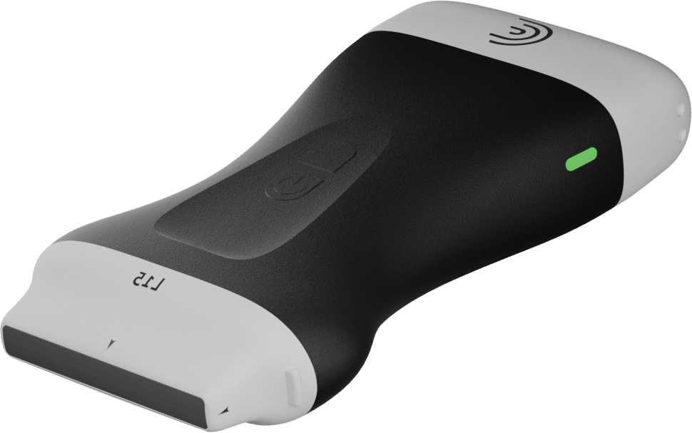
Dr. Rowland-Warmann uses the L20 Ultra-High Frequency Linear Scanner to visualize anatomy prior to filler placement each and every time, to ensure safety and the best results for her patients.
Interested in learning more about Clarius for Aesthetic Procedures?
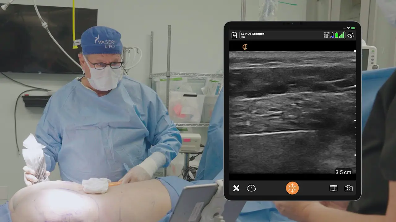
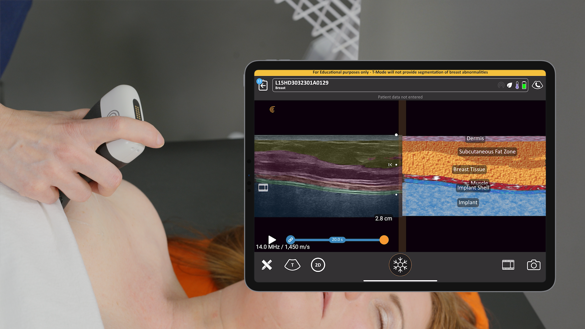
More aesthetic clinicians like Dr. Rowland-Warmann are using Clarius high-definition wireless ultrasound to ensure safer procedures and better results. Clarius is now more affordable and easier to use than traditional ultrasound systems. The Clarius L20 HD3 is the world’s first high-frequency handheld scanner, with exceptional superficial imaging and specialized for facial aesthetics.
Contact us today or request an ultrasound demo to see how high-definition imaging can help you improve safety and deliver consistent patient outcomes in your practice.
