Clarius Ultrasound for Clinical Research

Clarius ultrasound tools for research can be purchased with any scanner. They provide access to raw data collected internally and custom software for real-time analysis.

  • Mobile solution for clinical research and data acquisition
  • Multiple raw data formats
  • Streaming API and 3D positional information
  • Enabled with HIPPA-compliant Clarius Cloud for simple and powerful data management
    ultrasound-research-device

Quality Controlled Data Acquisition with AI Tools

With fully automated imaging workflows, Clarius scanners require minimal user intervention, making them ideal for large clinical studies that require operator independence and reproducible results. Through our innovative AI tools for automated TGC and parameter adjustment, study coordinators can focus on the clinical data without worrying about discrepancies from operator variance.

Easy Data Analysis Anywhere

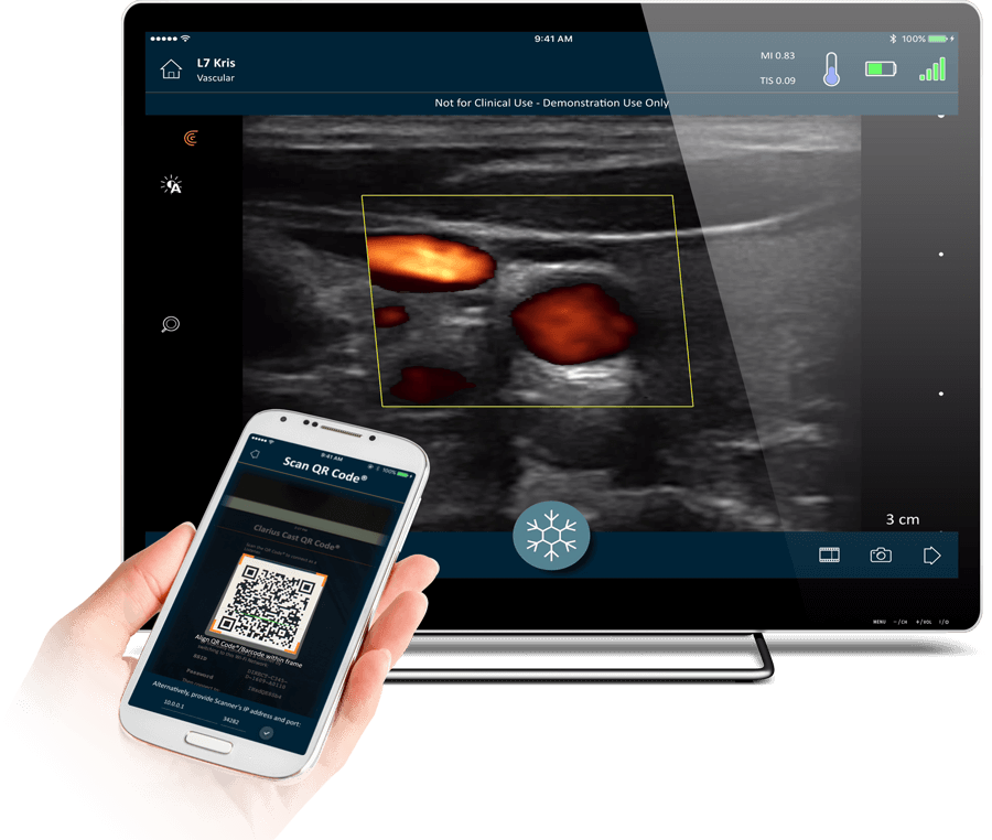
Thanks for the HIPAA-compliant Clarius Cloud, data analysis for clinical trials spanning global institutions is easy. All images and raw data are securely uploaded to the Clarius Cloud where additional measurement, analysis, and reporting can be performed. The data is available for analysis through the portal anywhere in the world with Internet access.

Clinical Indications for Research

Clarius scanners for clinical research collect images and raw data for analysis and measuring physiological parameters. Examples of clinical areas for research include: IMT and FMD for vascular health, aortic measurements for AAA screening, the use of power doppler for arthritis, or attenuation measurements for liver health.

The Raw Data Package allows users to collect raw envelope (B-mode), IQ, and RF signals from the scanner. When raw data is acquired by the user, it is automatically tagged with the corresponding image or cine clip captured during clinical operation.

Data Provided
  • Pre-scan converted greyscale with a range of 256 shades of grey.
  • Demodulated IQ in signed 32-bit pairs.
  • Beamformed RF in signed 16 bit format that has been digitized at 14-bits (pre-beamformed RF signals are not available on Clarius scanners).
Transfer
  • Downloaded into the Clarius App after the image is frozen by capturing a single image or cine clip.
  • Data transfers from the scanner to the App may take up to 30 seconds, depending on the buffer size collected. During this time, real-time scanning is not permitted.
  • Raw data is automatically sent to the Clarius Cloud (with proper Internet connection) once an exam has been completed.
Viewport
  • All data is acquired from the visible imaging sector.
Data Rates
  • When collecting only B and IQ data, the raw data is collected at the native imaging frame-rate. When additionally collecting RF data, the frame rate is reduced by 15 to 20%.
Buffering
  • Up to 30 seconds of envelope data, and up to 15 seconds of IQ and RF data (dependent on the scanner model and mode of operation).
Storage
  • Raw data is tagged and stored alongside the captured image or cine clip.
  • On Android devices, users can export raw data onto the mobile device storage once the exam has been completed.

The 3D Positional Data Package allows users to collect the 9DOF data from the inertial measurement unit (IMU) embedded on each Clarius scanner.

Data Provided
  • Gyroscope information from X, Y, and Z axes
  • Accelerometer information from X, Y, and Z axes
  • Magnetometer information from X, Y, and Z axes
  • Precise timestamp to help correlate IMU data with the ultrasound images.
Transfer
  • IMU data is automatically transferred (with proper Internet connection) to Clarius Cloud with the ultrasound images once the exam has been completed.
Data Rates
  • The pre-programmed IMU collection rate is 200 Hz, as imaging typically runs at 15 to 30 FPS, and each frame is tagged with multiple IMU data points.
Buffering
  • IMU data is collected for the entire duration of the scan, where the maximum cine buffer is programmed for 30 seconds.
Storage
  • IMU data is tagged and stored alongside the captured image or cine clip.
  • With the Raw Data package, IMU data will also be stored in the raw data files that are collected from the scanner.

The Listener API works like Clarius Cast, where multiple devices can view an ultrasound scan in real-time. The Listener API enables users to build custom software to receive a live ultrasound stream from a Scanner already connected from a Clarius App.

Data Provided
  • Real-time streaming of scan-converted B mode and Color Doppler images
  • Raw data such as IQ, RF, as well as IMU data cannot be streamed through the API.
Connectivity
  • The API must be running on a device that is already connected to the scanner’s WiFi Direct. Alternatively, all devices can be connected to a local WiFi router.
  • The API must be programmed with the Scanner’s IP address and the UDP streaming port in order to make a connection. This information can programmed to be static to minimize program setup.
Data Rates
  • Data rates on WiFi vary. In most cases, the Listener API will be able to receive ultrasound images at the native framerate with little to no latency.
Buffering and Storage
  • It is up to the software written using the Listener API to display, buffer, and store images should that functionality be desired.