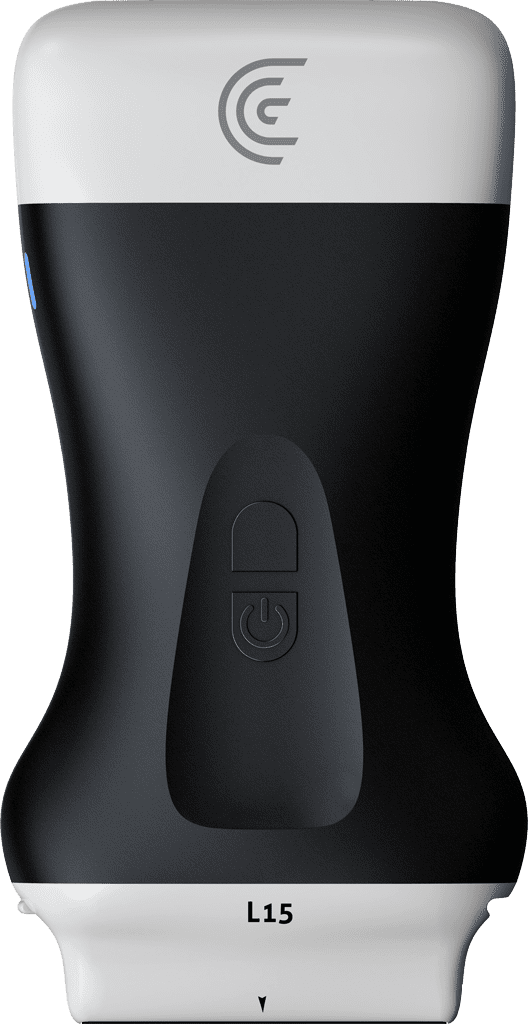
In this Clarius Classroom tutorial, physical therapist C.J. Miller from Frontline Function demonstrates how to use the Clarius L15 scanner to perform side-to-side comparisons on a patient’s anterior knee.
Key Highlights
Anatomical Navigation: Miller places the probe in the long axis to visualize the patella on the left side of the screen and the tibial tuberosity on the right, linked clearly by the patellar tendon.
Identifying Abnormalities: While scanning the width of the tendon’s insertion, Miller identifies significant cortical changes on the bone at the tibial tuberosity.
Split-Screen Functionality: To determine if the findings are normal for this specific patient, Miller freezes the image at the peak of the abnormality and utilizes the ultrasound’s split-screen mode.
Confirming Pathology: By scanning the opposite (right) knee in the same area, Miller reveals a much smoother bone surface with far fewer cortical abnormalities, confirming the left side’s pathology.
Clinical and Educational Value: Real-time side-to-side comparisons are highlighted as a powerful tool to help practitioners confirm abnormalities and help patients visually understand the source of their pain.