


Lip augmentation remains a staple in aesthetic medicine for men and women. But concerns over complications—particularly vascular events—are on the rise. Renowned medical aesthetic expert Dr. MJ Rowland-Warmann, founder and lead clinician at Smileworks in Liverpool, advocates for the use of ultrasound as the “most groundbreaking development in aesthetic medicine since the innovation of filler.”
Our free on-demand webinar with Dr. MJ on ultrasound-guided lip fillers is popular among aesthetic physicians interested in learning safer techniques for lip augmentation. We invite you to watch it at your convenience. Scroll on for clinical highlights and Dr. MJ’s detailed video demonstrations.
Beyond Blind Injection: Mastering Lip Fillers with Ultrasound Guidance
Ultrasound is known to provide essential information about sub-dermal anatomy, enabling practitioners to avoid the labial arteries and plan safer, more predictable injections. We’ve compiled this article as a guide to Dr. Rowland-Warmann’s expert techniques, providing details on how to utilize ultrasound for safer and more effective lip augmentation.
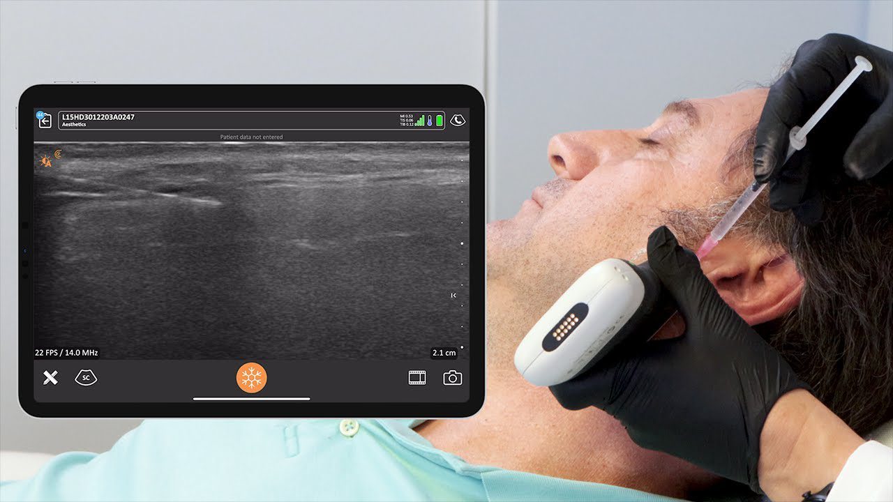
Dr. MJ integrates ultrasound throughout the filler process—before, during, and after treatment.
- Pre-treatment: To identify high-risk anatomy, such as the variable positions of the labial arteries.
- During treatment: Using real-time guidance to precisely place filler, ensuring it is away from critical vascular structures.
- Post-treatment: Checking filler positioning, monitoring product over time, and reassuring the patient of a job well done.
- Complication Management: It is revolutionary for the diagnosis and management of aesthetic complications, allowing for quick and effective resolution.
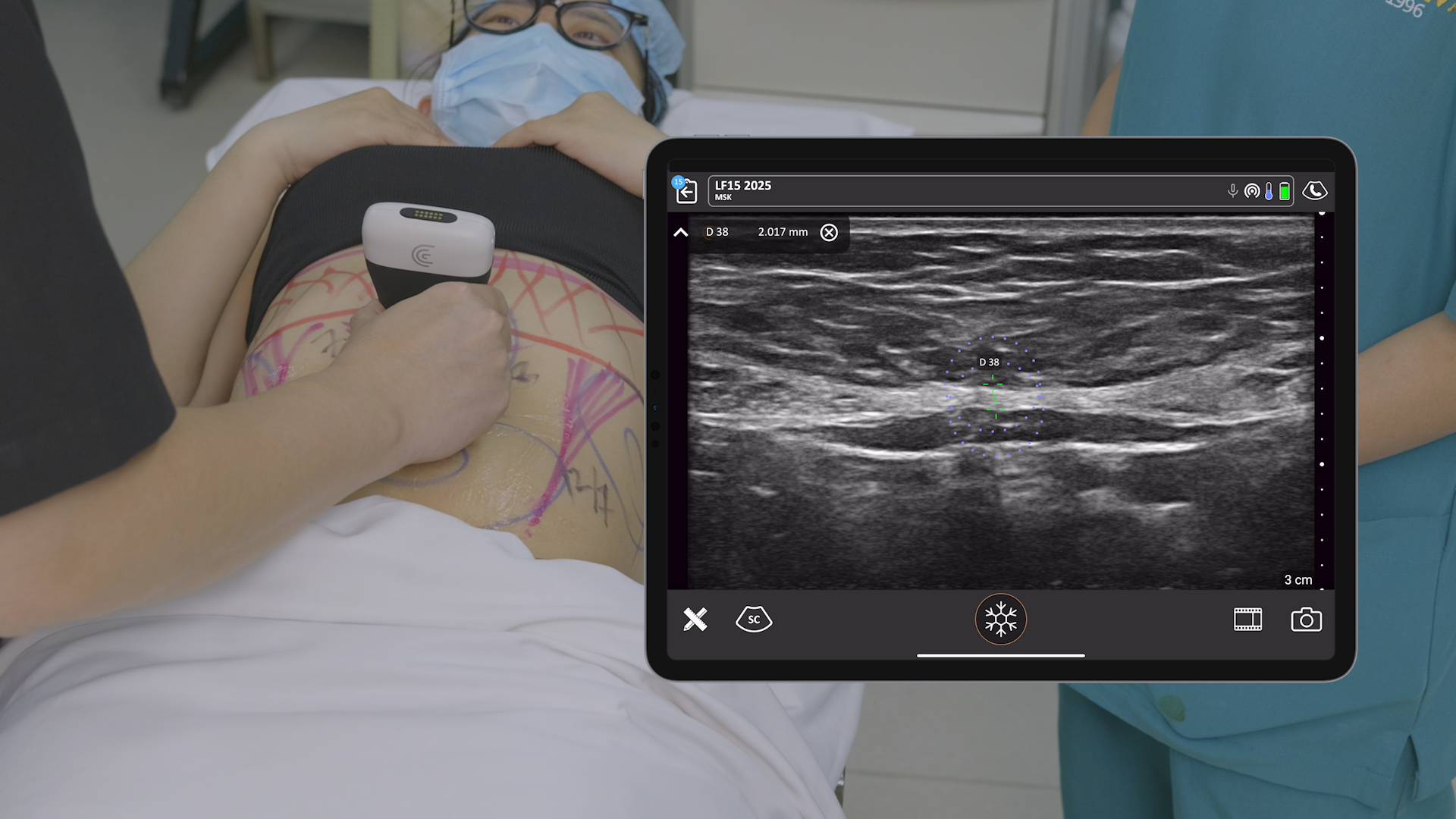
Essential Scanning: Finding the Labial Arteries and Filler
Understanding lip anatomy is paramount, as the placement and course of the labial arteries are highly variable.
Dr. MJ’s Go-To Scanning Technique
Dr. Rowland-Warmann’s favorite technique for a quick, efficient assessment is the vertical transducer angulation.
- This view provides the best overview of the relationship between the lip layers and the arteries.
- Scan the lateral and central parts in each quadrant (upper and lower lip) to get the necessary overview.
- Assessing in cross-section determines the vessel location and depth, which informs the injection technique.
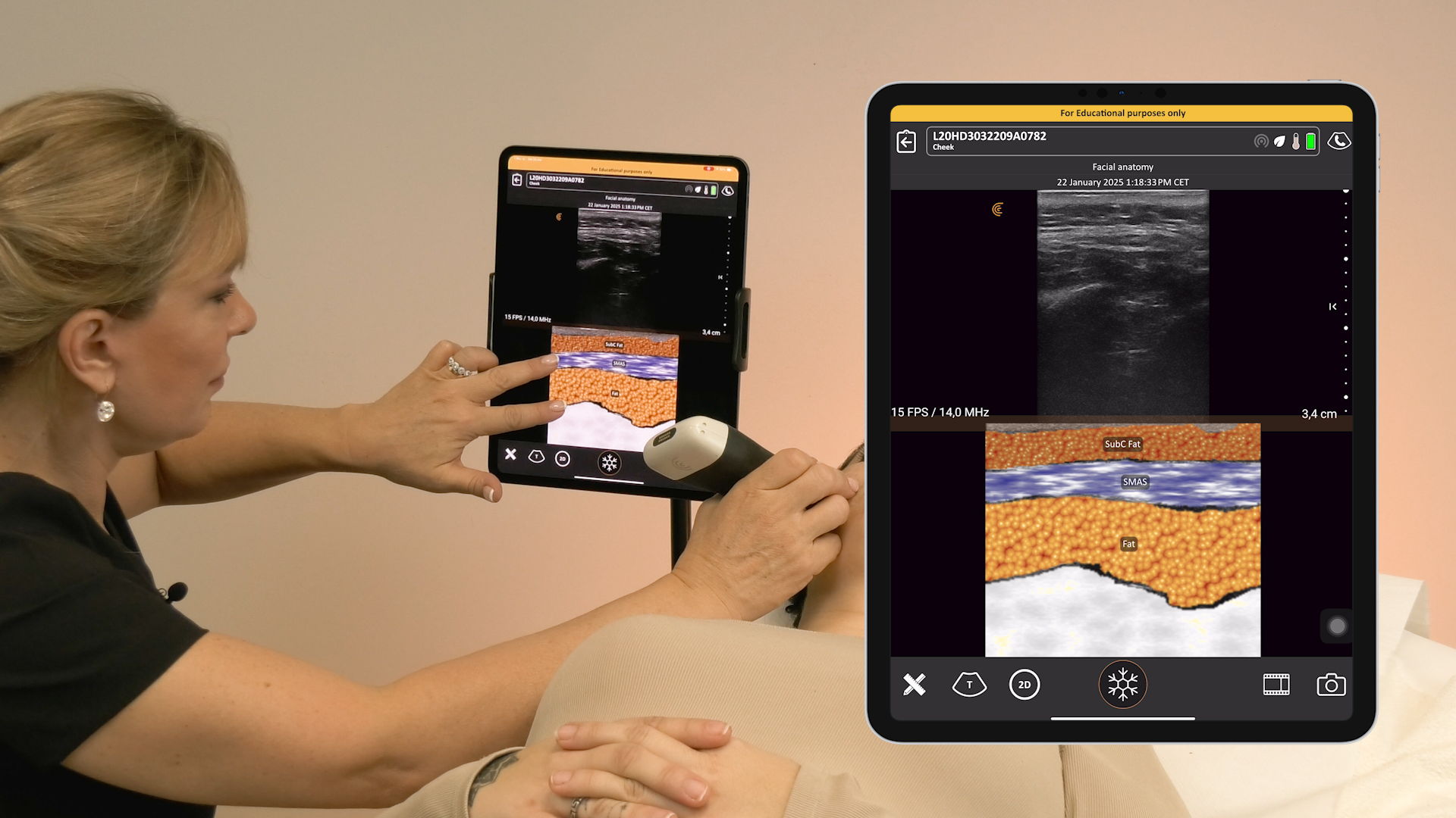
Key Anatomical Insights
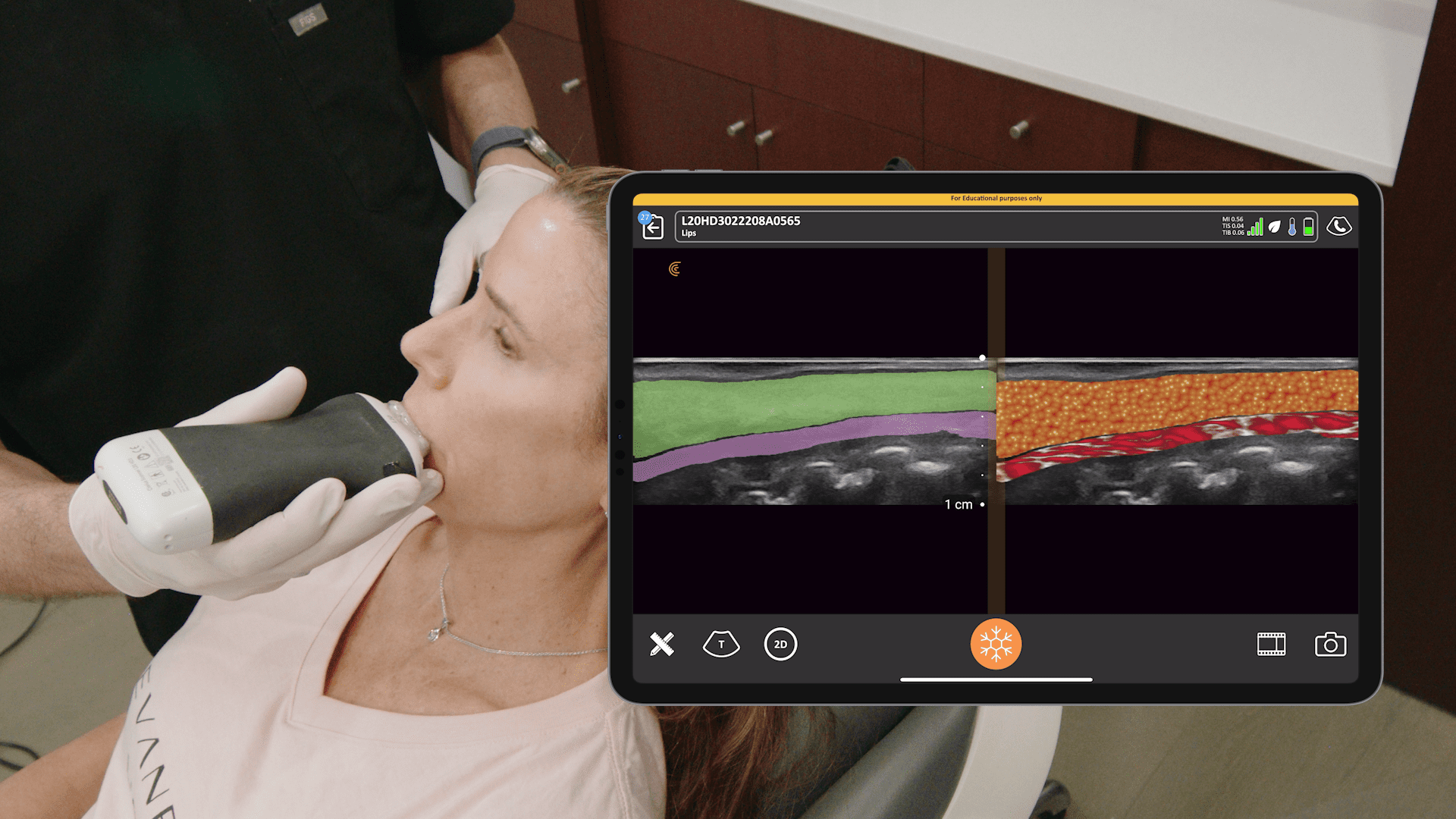
- Lip Layers: External to internal, the lip has five tissue layers: skin, subcutaneous fat, orbicularis oris, submucosal fat, and oral mucosa.
- Target Layer: The aim for lip filler is the subcutaneous layer of the dry red lip (vermilion). The recommendation is to inject subcutaneously with a depth of no more than three millimeters.
- Artery Location Variability: Studies show that labial arteries can be submucosal (most common, e.g., 58.5% in one study), intramuscular (e.g., 36.2%), or subcutaneous (e.g., 5.3%).
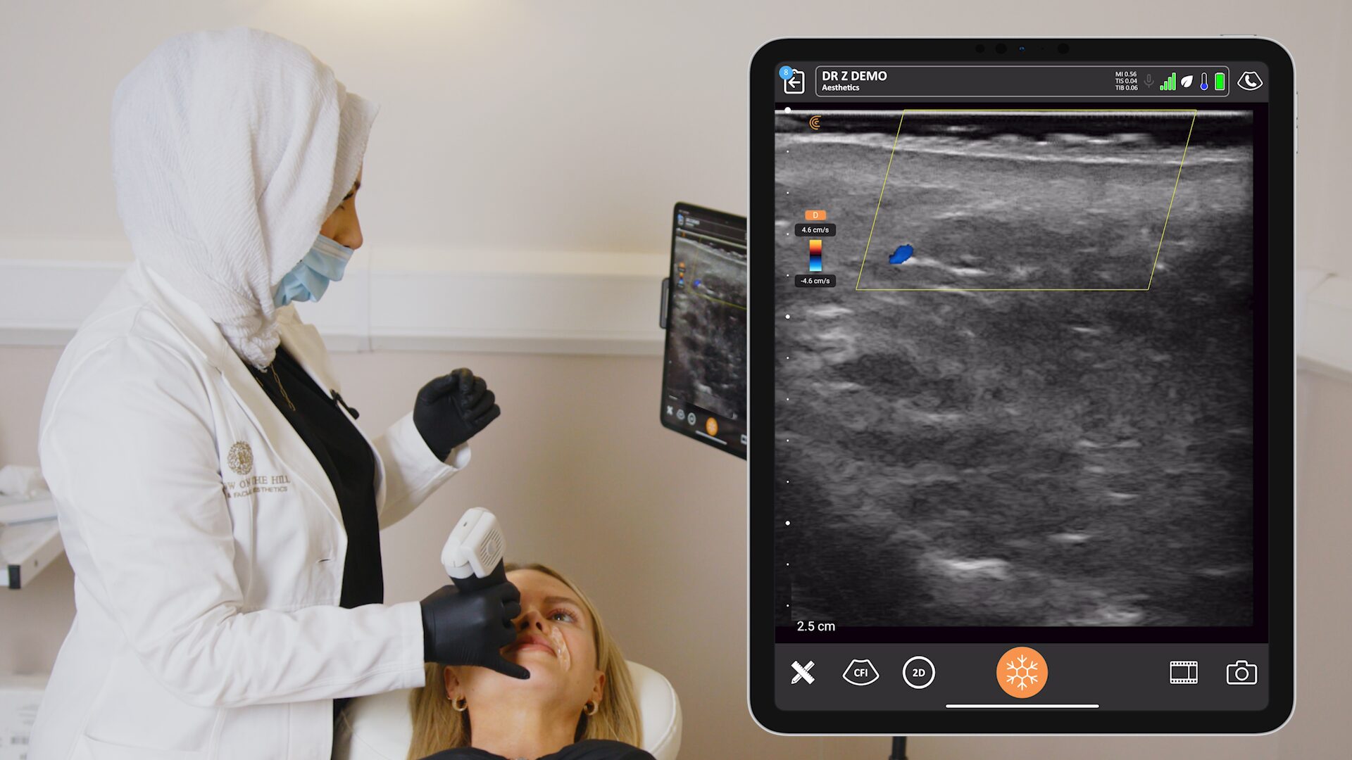
- Identifying Filler: Hyaluronic acid (HA) filler in B-mode appears as anechoic (black) oval deposits, sometimes exhibiting posterior acoustic enhancement (little white saucer shapes deep to the structure).
- Differentiating Filler from Vessels: HA filler deposits do not exhibit flow when Color Doppler is applied, which helps to distinguish them from vessels.
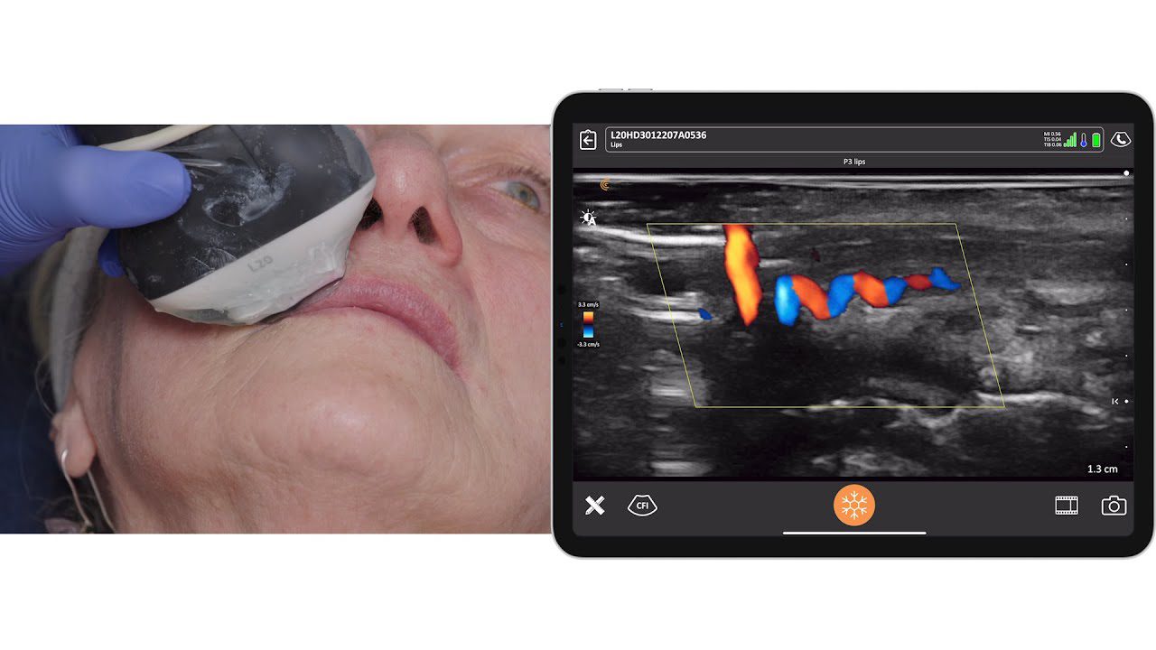
Video: Vessel Mapping in the Lips
Watch this video to see how Dr. MJ Rowland-Warmann confirms the position and course of the labial arteries in the lips using the Clarius L20 HD3.
Planning the Injection: Needle vs. Cannula
The location of the labial artery determines the safest injection method.
| Artery Location | Recommended Injection Tool | Rationale |
| Submucosal Plane | Needle | Safe to inject superficially in the subcutaneous plane, away from the artery. Dr. MJ uses a subcutaneous linear threading technique for precision. |
| Subcutaneous Plane | Guided Cannula Approach | This is the exception (around 5% incidence). Direct visualization is key to avoid vessel injury. |
Safety Alert: Techniques that involve inserting a needle perpendicular to the vermilion border (like vertical strands or ‘Russian lips’) significantly increase the risk of vascular adverse events due to the needle tip’s proximity to the labial artery.
4-Minute Video: Ultrasound-Guided Lip Filler Treatment
In this video, Dr. MJ provides an overview of the tissue layers of the lip, including the identification of vascularity and existing filler, before guiding her needle to dissolve HA filler deposits. She uses her Clarius L20 HD3.
Fixing Complications: Guided Dissolving
When filler is in the wrong place, such as in cases of filler spread, the most effective solution is to dissolve it and start again.
- Unguided vs. Guided: Ultrasound-guided hyaluronidase deployment is far superior, resulting in improvement in 100% of cases (compared to 7.7% for unguided) in the management of vascular adverse events.
- Benefits: Guided dissolving utilizes less hyaluronidase, is more precise, and results in quicker improvement.
- Technique: Dr. Rowland-Warmann uses an in-plane technique (needle in line with the transducer) to guide a needle (often 27 gauge) directly to the HA deposit and inject a small, targeted amount of hyaluronidase.
- Hypervascularity: Lips with a lot of previous filler often exhibit hypervascularity (increased blood flow and vessel proliferation) on Doppler, which is an irritant effect of the filler on the vessels.
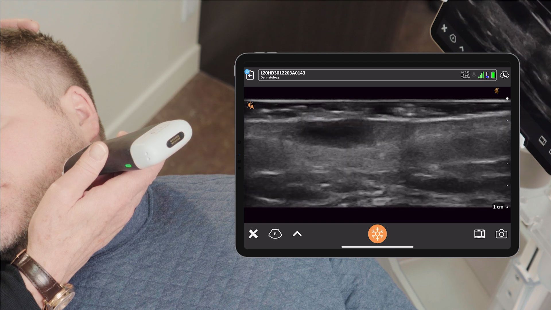
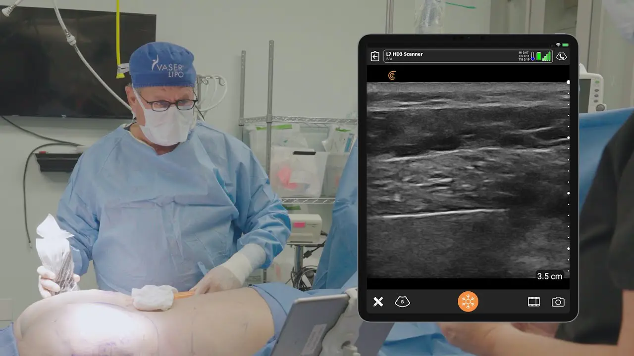
Why Clarius Ultrasound is a Popular Choice for Aesthetic Physicians
The miniaturization and reduced cost of handheld ultrasound devices are making high-quality ultrasound scanning accessible to aesthetic professionals.
The Clarius L20 HD3 is the world’s only ultra-high-frequency (8–20 MHz) wireless scanner with specialized software for aesthetics, providing high-resolution superficial imaging up to a 4 cm depth, ideal for facial anatomy.
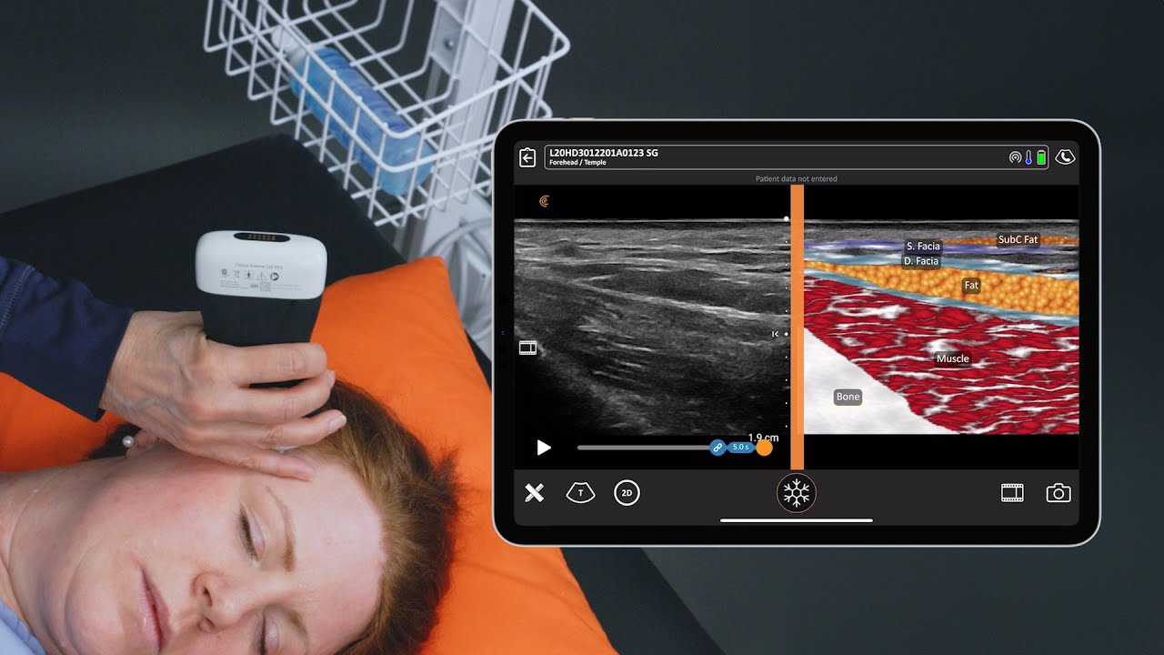
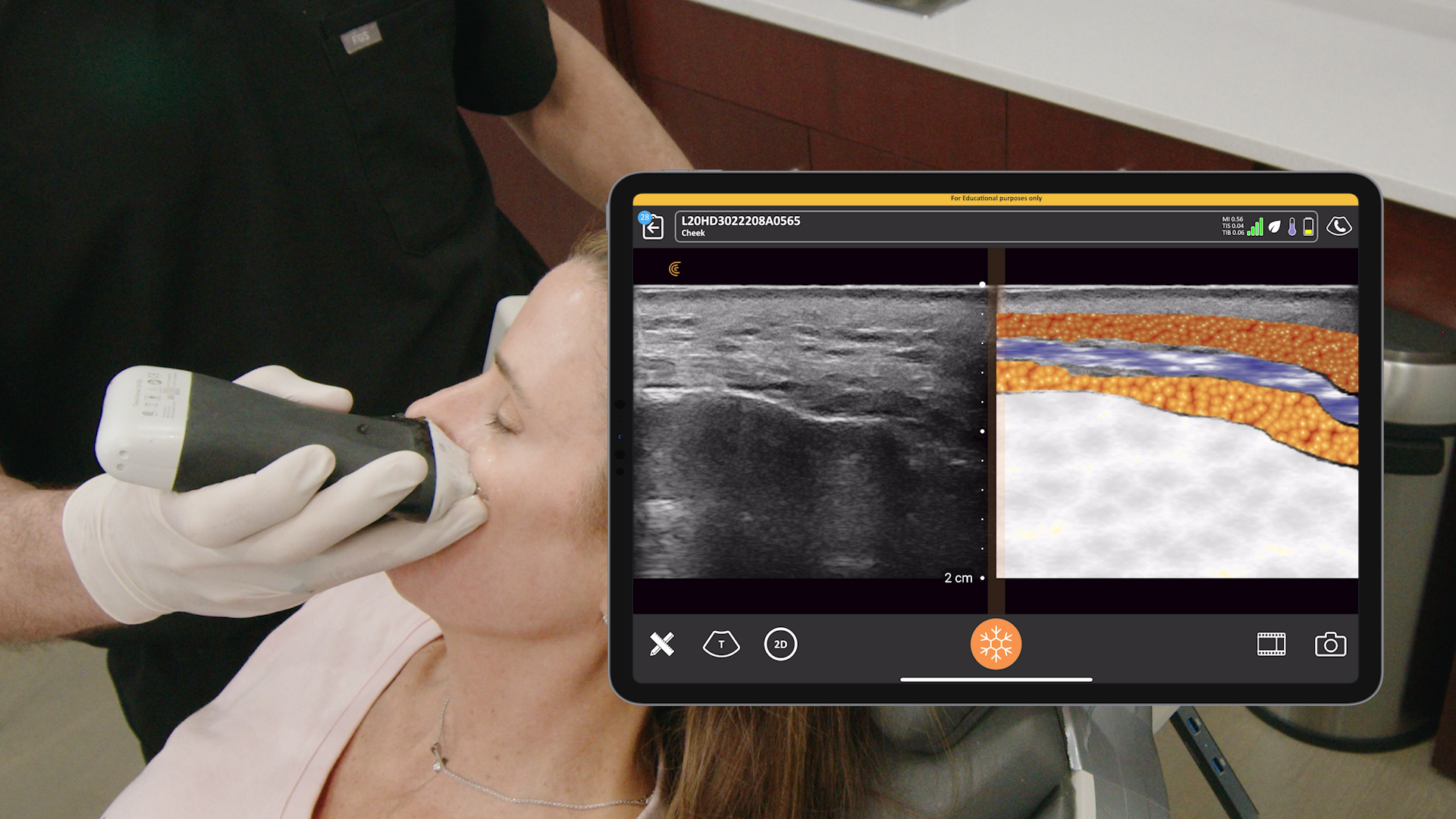
Clarius Intelligence Features:
- T-Mode: Automatically colors in tissues to distinguish layers (like fat and muscle) and provides on-screen anatomical labels, making it easier to distinguish and learn ultrasound anatomy.
- Vessel Depth AI and aesthetic presets further automate workflows and optimize imaging parameters.
- Needle Enhance: Boosts confidence and accuracy by providing clear visualization of the needle during procedures.
Portability and Ease of Use: The scanner is ultra-portable, wireless, and works with an easy-to-use app for iOS and Android devices, streamlining workflows with simple controls and automatically delivering optimal imaging.
Clinical Efficacy: An independent study at the University of Southern California ranked the Clarius L20 HD3 as having the highest image quality for filler and orbital imaging among several point-of-care ultrasound devices.
Learn more about Clarius AI-powered ultrasound on the aesthetics specialty page. Or contact us for a personalized virtual demonstration.
About Dr. MJ Rowland-Warmann
Dr. MJ Rowland-Warmann is the founder of the Smileworks Aesthetic Training HUB, which offers both Foundation and Advanced Facial Ultrasound Training courses. These courses combine CPD-accredited online learning modules with intimate, hands-on training sessions to help experienced injectors integrate ultrasound, refine their precision, and manage complications with greater confidence and control.


