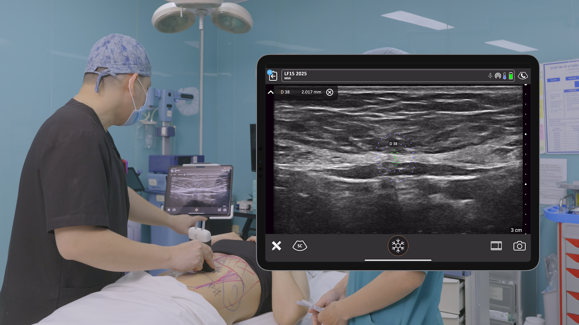
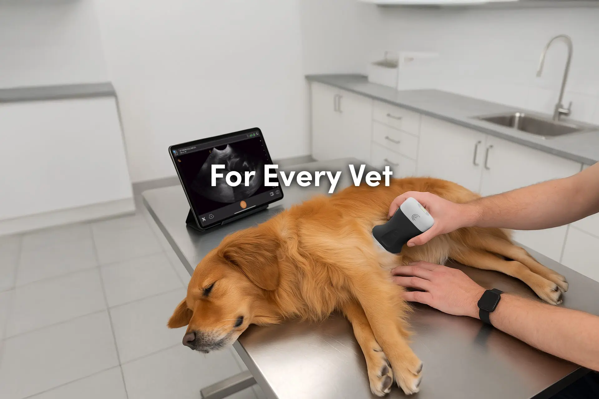
Once primarily a tool used by radiologists, ultrasound systems are now available and commonly used by leading medical specialists who offer treatments for every part of the human body. Recognizing the rising popularity of aesthetic procedures and the availability of high frequency ultrasound systems capable of imaging the skin, the European Federation of Societies for Ultrasound in Medicine and Biology (EFSUMB) has released a detailed position statement on the use of dermatologic ultrasound.
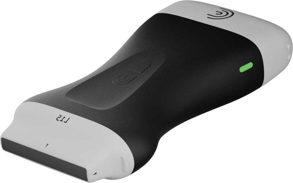
As Clarius handheld ultrasound is the leading choice for aesthetic clinicians, we’ve put together a quick capsule of the guidelines for ultrasound use related to aesthetic medicine. Read on for highlights and to learn why the Clarius L20 HD3 and the Clarius L15 HD3 are the only handheld ultrasound scanners that meet EFSUMB guidelines.
Minimum Transducer Frequency Should Be 15 MHz for Aesthetic Procedures
Due to limited computing power, it hasn’t always been possible to use ultrasound to examine superficial structures like the skin. And when high frequency ultrasound transducers (>20Mhz) were first introduced 20 years ago, they were only used by institutions that could afford the high cost of acquiring premium ultrasound systems, which were often dedicated to clinical research.
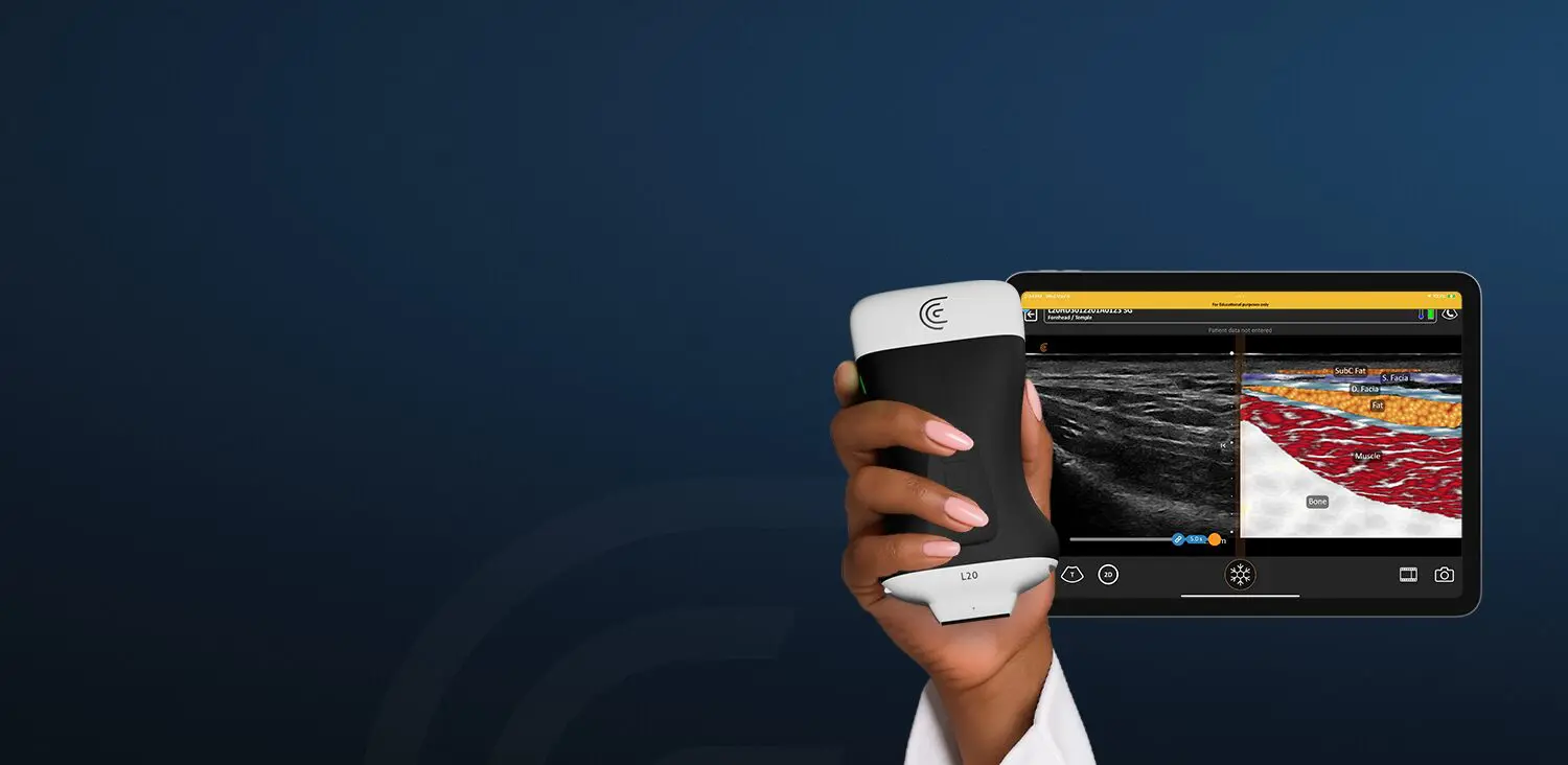
Now, high frequency transducers are more widely available and EFSUMB is recommending a minimum standard of a 15 MHz transducer for dermatologic ultrasound. While there are numerous cart-based and lap-top systems with high frequency transducers on the market, the Clarius L20 HD3 is the only handheld ultrasound system that offers a frequency range of 8-20 Mhz. With a price that is a tenth of the cost of a traditional ultrasound system, Clarius ultrasound has become a popular choice for aesthetic dermatologic ultrasound.
Dr. Ines Verner, an Aesthetic Dermatologist based in Tel-Aviv, has been using Clarius ultrasound for more than a year and has this to say: “Clarius is a good fit for aesthetics, because it’s easy to use, the image quality is good. We can really see what we need to see. I think the new Clarius L20 HD3 is a very good technology for the aesthetic clinic. The transducer head is quite small, so we can use it in different areas of the face.”
Watch our video interview with Dr. Verner to learn why she thinks ultrasound is the new breakthrough in Aesthetic Dermatology internationally.
Color Doppler/Power Doppler and Pulsed Spectral Doppler Are Recommended To Detect Vascular Anatomy
To detect vascular anomaly and to detect inflammation, the EFSUMB steering committee also recommends that all ultrasound exams include color, power or spectral Doppler.
You can see the Clarius L20 HD3 in action in this video where Dr. Ines Verner uses Color Doppler to visualize the location and measure the depth of supratrochlear and supraorbital arteries.
Ultrasound Is Recommended for Evaluating Facial Anatomical Structures
Given the known complexity and variability of anatomical structures in the face, the steering committee considers ultrasound to be a valuable tool for planning techniques prior to procedures and for managing care and possible complications after the procedure.
Dr. Steven F. Weiner, a board-certified head, neck and facial plastic surgeon, is an avid user of ultrasound for facial mapping. Watch our 1-hour free webinar to learn Dr. Weiner’s techniques for clearly visualizing facial vascular network in minutes to eliminate risk and keep patients safe.

Ultrasound in Facial Aesthetics: Vascular Mapping, Evaluating, Fillers and Complications
Dr. Weiner explores:
- The real dangers of blind injections and how to eliminate them
- Vascular mapping to confirm the location, size and depth of arteries
- How to identify and characterize previously placed fillers
- Guiding injections to dissolve HA fillers and placing steroids in granulomas
Ultrasound Is Recommended To Detect Common Types of Fillers, Implants and Related Complications
Clarius users report that many patients don’t have records of the types of cosmetic treatments they have received over time. So, we were not surprised to see that the WFSUMB Steering Committee recommends using ultrasound to identify common types of cosmetic fillers and implants and also to manage complications.
Dr. Stella Desyatnikova, MD, a double board-certified plastic surgeon, recalls a time when she was called upon to reverse a vascular occlusion that occurred following a filler injection to the lips and chin.
Using her Clarius ultrasound scanner, she found an area with abnormal blood flow in the chin where a small lump of filler was putting pressure on the arteries.
We injected the lump of filler with only half a vial of hyaluronidase under ultrasound guidance. Immediately, skin appearance and capillary refill were improved and blood flow on ultrasound dramatically improved as well,” Dr. Stella explains. “The patient’s facial skin looked good the next day and she is now completely back to normal.”


Percutaneous Ultrasound Guidance Is Recommended for Precision and to Decrease Complications
The WFUSMB committee recognizes the benefits of using percutaneous ultrasound guidance to improve the precision of injections, drainage procedures, to decrease adverse reactions and complications.
Dr. Marc Salzman, a double-board certified plastic surgeon, is no stranger to using ultrasound at his practice. We invited Dr. Salzman to present a webinar to teach ultrasound best practices to improve breast augmentation outcomes. See the Clarius L15 HD3 in action during the one-hour webinar where Dr. Salzman teaches how to detect, measure, and monitor seromas and ALCLs. Dr. Salzman shows how ultrasound allows you to visualize your needle under ultrasound guidance to safely and accurately aspirate the fluid without sticking the implant.
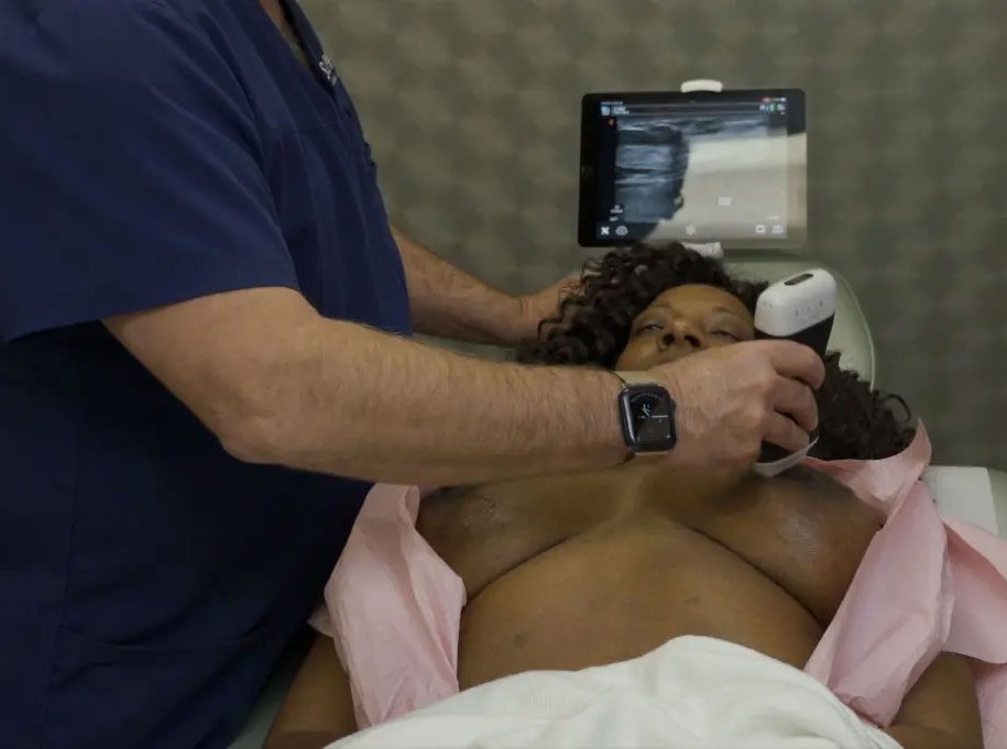
[FREE WEBINAR] Ultrasound Essentials for Breast Plastic Surgery: Visualizing Silent Ruptures, Seromas, and PECS Blocks
Dr. Salzman explores:
- Use a breast scanning protocol and characterize existing implants
- Accurately detect silent ruptures with ultrasound, a case study
- Use ultrasound to confirm that an implant is upside down, a case study
- Perform accurate Pectoralis Blocks using ultrasound-guided techniques, a case study
- Diagnose and safely guide aspiration of breast seromas under ultrasound
Ultrasound Is Recommended for Evaluation of Plastic Surgery Procedures and Complications
The WFUSMB positioning statement states: “Ultrasound can detect complications of cosmetic or plastic surgery procedures, such as dermal and hypodermal edema, lymphedema, seromas, hematomas, abscesses, fistulous tracts, fat necrosis, granulomas and loose sutures.”
In the ultrasound clip below, captured with a Clarius scanner, Dr. Marc Salzman aspirates a seroma. The red dots are along the needle. You see the needle come into the cavity and you can watch the black, or the hypoechogenic fluid, slowly get closer and closer so that the black is gone, and the regular, lighter-colored soft tissues are coalescing.

A Written Post Exam Report with High Quality Ultrasound Images Must Be Provided
The WFUSMB committee recommends creating a written report with ultrasound images that document that anatomy captured by ultrasound, ideally on a computer-based system.
Another reason that Clarius ultrasound scanners are an ideal fit for practitioners looking for an ultrasound system that meets EFUSMB guidelines, is the Clarius Cloud exam management portal, which often tops the list of reasons our users love Clarius.
I’ve tried all the handheld ultrasound systems. The coolest thing about Clarius is the cloud system. It’s miles ahead of all other machines,” says ultrasound educator Chris Myers. “I love the fact that you just go on your computer and your scans are there. You can write reports, label the images and simply email them off to colleagues and referrers. I’ve also used it for teaching online during lockdown.”
Theoretical and Practical Educational Courses Are Recommended
Since dermatologic ultrasound is relatively new, the WFUSMB committee emphasizes the important of practical and theoretical courses to improve ultrasound proficiency.
Many Clarius users are leading educators in the field and are regular contributors to our education program. For example….
Book a Personal Demonstration of the Clarius Wireless Ultrasound Scanner for Dermatologic Ultrasound
We would be delighted to discuss how Clarius ultrasound can help you improve safety and patient outcomes at your practice. Contact us today or request an ultrasound demo with a Clarius expert in your region.
