
When Dr. Steven Weiner started using ultrasound guidance to improve outcomes and reduce complications resulting from facial cosmetic procedures, he didn’t realize that he’d soon become an internationally renowned speaker and educator.
Not that long ago, he flew to Amsterdam to learn from Dr. Leonie Schelke, who has been using ultrasound at her clinic to treat dermal filler complications for more than ten years. Following three days of training and a year of using ultrasound for safe cosmetic procedures at his clinic, Dr. Weiner says he injects with confidence using ultrasound guidance. What’s more, he’s now teaching ultrasound best practices to improve patient safety and deliver consistent results with aesthetic procedures.
If you know your anatomy and you know the vascular structures and you know where you’re placing your filler, then the patients are obviously going to do better,” he explains. “And God forbid, if there is a vascular complication, you’ll be able to identify it rather quickly and inject the pocket of filler using ultrasound guidance and eliminate the vascular occlusion very easily.”
Treating a Nasal Occlusion With Ultrasound Guidance Reduced Hyaluronidase Units by 96%!
Dr. Weiner recounts an incident while training with Dr. Schelke that hit home for him. “Someone called into the clinic to ask for guidance on how to treat a patient with a nasal occlusion from an injection in the nose. Dr. Schelke said she could see the patient the next day and in the meantime suggested giving her 3,000 units of hyaluronidase into the nose.
When we saw the patient the following day, the patient was still occluded. Using ultrasound, we were able to clearly identify the areas of occlusion. We were then able to inject 125 units of hyaluronic acid under ultrasound guidance and instantly fix the problem.
Just the day before, 3,000 units hadn’t fixed the problem because it wasn’t injected in the right place. We injected 125 units under ultrasound guidance and fixed the problem. And, we were able to see immediate flow of the vessels that had been occluded. We did a follow-up evaluation the next day and the patient’s nose was normal. So that’s the power of ultrasound!”
It’s phenomenal that under ultrasound guidance, a 96% reduction in units of hyaluronidase (from 3,000 to just 125) was needed to successfully resolve the filler complication.
Ultrasound is the Gold Standard for Evaluating Fillers and Vascular Mapping
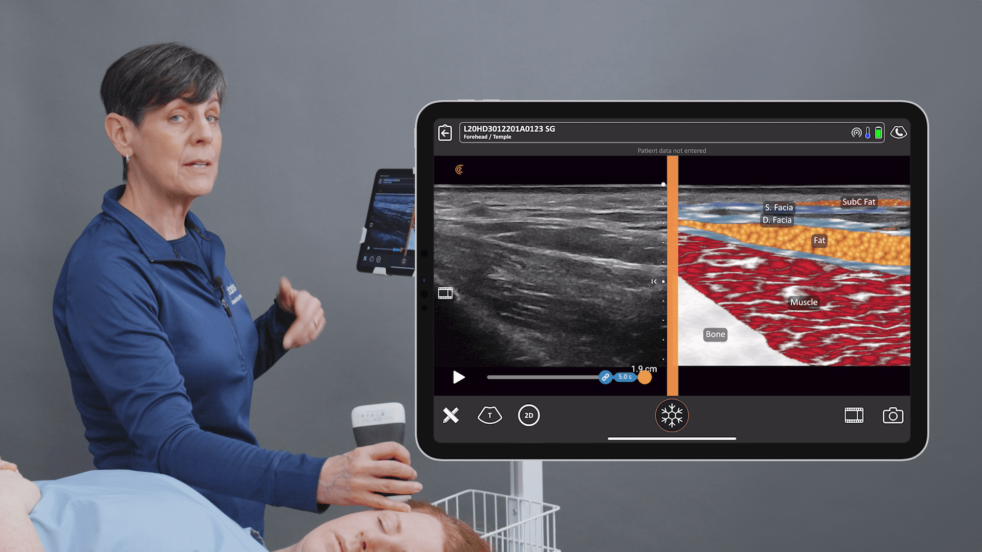
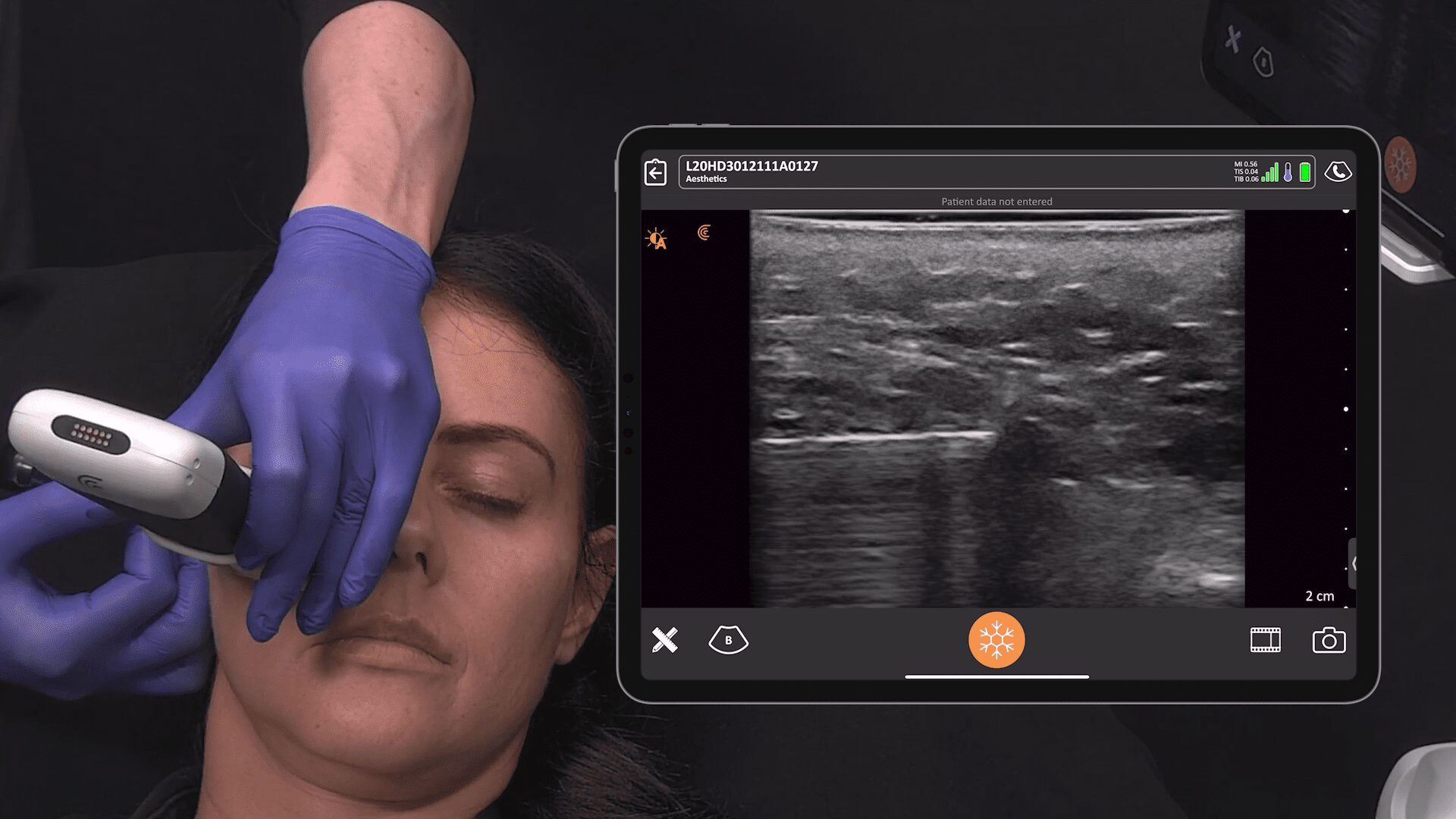
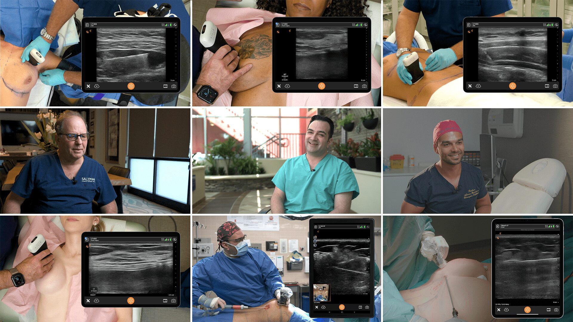
Dr. Weiner is a board-certified head, neck and facial plastic surgeon who practices at the Aesthetic Clinique in Florida, which he established 15 years ago. He uses the Clarius L20 high-frequency ultrasound scanner, which he considers ideal for facial aesthetic procedures with its ability to provide high-definition images to a maximum depth of four centimeters.
“Ultrasound is the gold standard for evaluating patients, particularly with filler complications,” he says. “But it’s better to be pre-emptive by evaluating vascular structures using vascular mapping in advance to avoid these structures when you’re doing the injection.”
Dissolving HA Fillers Requires Ultrasound to Guide the Needle Placement Within the Filler Pocket
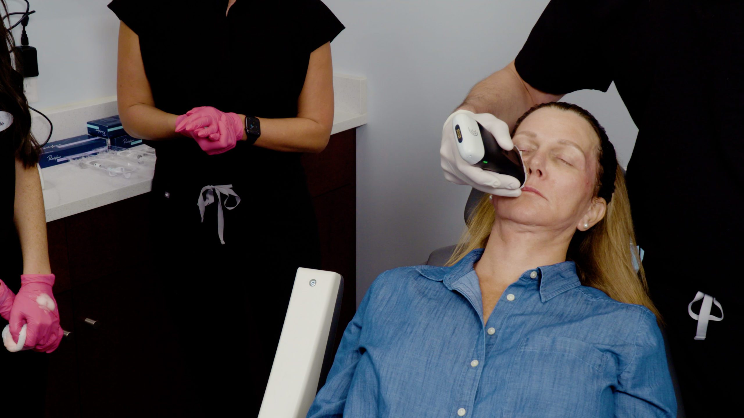
In the following Clarius Classroom video, Dr. Weiner’s assistant and a nurse use ultrasound guidance to dissolve an HA filler on Dr. Weiner himself.
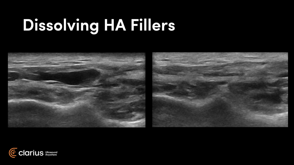
“These images you see here are of my cheek before and after. On the right is an image captured 24 hours after the hyaluronic injection. You can see it’s basically dissolved. This was done under ultrasound guidance.
I had previously tried injecting this area blindly without success. I find that fillers form a little cyst, or pseudocyst, with a little wall around them. That’s why patients come to me and say that they’ve been injected four, five, six times and they still have the nodule or swelling related to their prior HA.
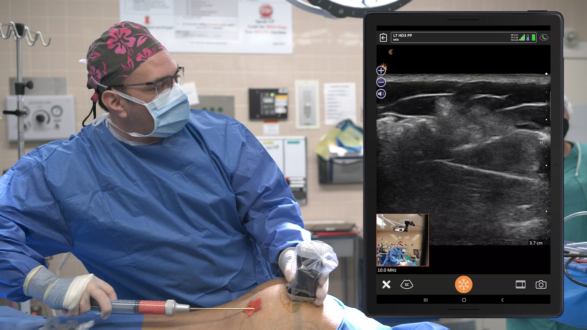
You can see in the video that we are actually within the pocket. You can see the cannula and you can see the resolution of the nodule before your eyes!
You can do this procedure by yourself on a patient with the canula in one hand and the scanner in the other, but it is a little easier with an assistant. It was a painless procedure. I just used lidocaine prior.”
To learn more about how to use ultrasound to minimize the risks and complications associated with dermal filler procedures from Dr. Weiner, we invite you to watch his on-demand webinar, Ultrasound in Facial Aesthetics: Vascular Mapping, Evaluating Fillers and Complications.
Or read this article: Why Handheld Ultrasound is Essential for Safe, Consistent Dermal Fillers Outcomes.
With the miniaturization of high-definition ultrasound and advances in artificial intelligence, it’s now easy and affordable to add Clarius HD to your aesthetics practice. Contact us today or request an ultrasound demo to see the difference ultra-high-definition imaging can make to improve safety and patient outcomes in your aesthetic practice.


