Aula Clarius

Clarius T-Mode Abdomen Vet

Shelley Guenther, CRGS, CRCS

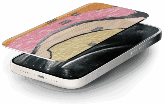
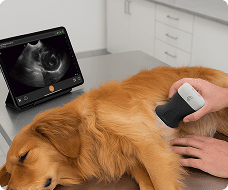
In this instructional video, Shelly and her dog, Squish, demonstrate how to use the T-Mode Abdomen Vet feature on a Clarius veterinary scanner. Designed as an educational tool to help new users recognize ultrasound anatomy, T-Mode automatically highlights and labels internal structures in real time.

Key Highlights

Scanner Setup: Shelly begins by using the abdominal preset and selecting the small animal option, noting that users can easily tap to adjust footprint sizes and depth for larger dogs.

Spleen Identification: Upon placing the scanner on Squish’s abdomen and activating the «T» button at the bottom of the screen, the software instantly generates a graphical representation of the anatomy, with live labels clearly identifying the spleen.

Gallbladder and Liver Evaluation: By pointing the scanner underneath the right rib cage, T-Mode dynamically identifies and labels the gallbladder and liver in real time.

Image Capture and Review: The demonstration highlights the ability to freeze the screen, scroll back to find the optimal frame, and capture an image—saving both the ultrasound image and its labels directly to the cloud.

Additional Applications: Shelly notes that T-Mode is also highly effective for identifying kidneys, making it a valuable feature for those navigating their ultrasound learning journey.

Specialties: Veterinary
Applications: Vet

00:00
–
01:01
Hi, I'm Shelly, and this is squish. And today we're going to tell you about T mode abdomen vet. So tea mode is an educational tool to help new users identify ultrasound anatomy. It can be challenging sometimes. So this is just a nice little tool to to help you along in your learning journey. So I'm using the vet scanner and I'm using the abdominal preset here.
And I've selected small animal. But you'll see here we've got different footprint sizes here. If the if we need more depth, if we have a bigger dog, we can just easily tap on those. So I'm going to place my scanner just on squishes abdomen here. And I haven't really done much. But what we're going to do here is just activate the T mode.
There's a T at the bottom of the screen here, and right away you're going to see, uh, a graphical representation of some anatomical structures in there. So I'm going to activate live labels here. And that's telling me that we're looking at the spleen. I can freeze my image
01:02
–
01:35
and take a picture and the image and the label will be stored to the cloud. So if we unfreeze and go back to 2D imaging here, let's, um, let's go a little bit over to the right girl. And I'm just pointing up underneath her right rib cage. So let's activate the T mode again. And we can see that we're the T mode is identifying the gallbladder and the liver here in real time.
So again I can freeze my image. I can send it back until I get the image that I like. And then we can store another image.
01:36
–
01:48
So the T mode is also really good for identifying the kidneys as well. So a great tool to help you get started in recognizing ultrasound anatomy. So have fun with the T mode.

Productos utilizados

Más clases