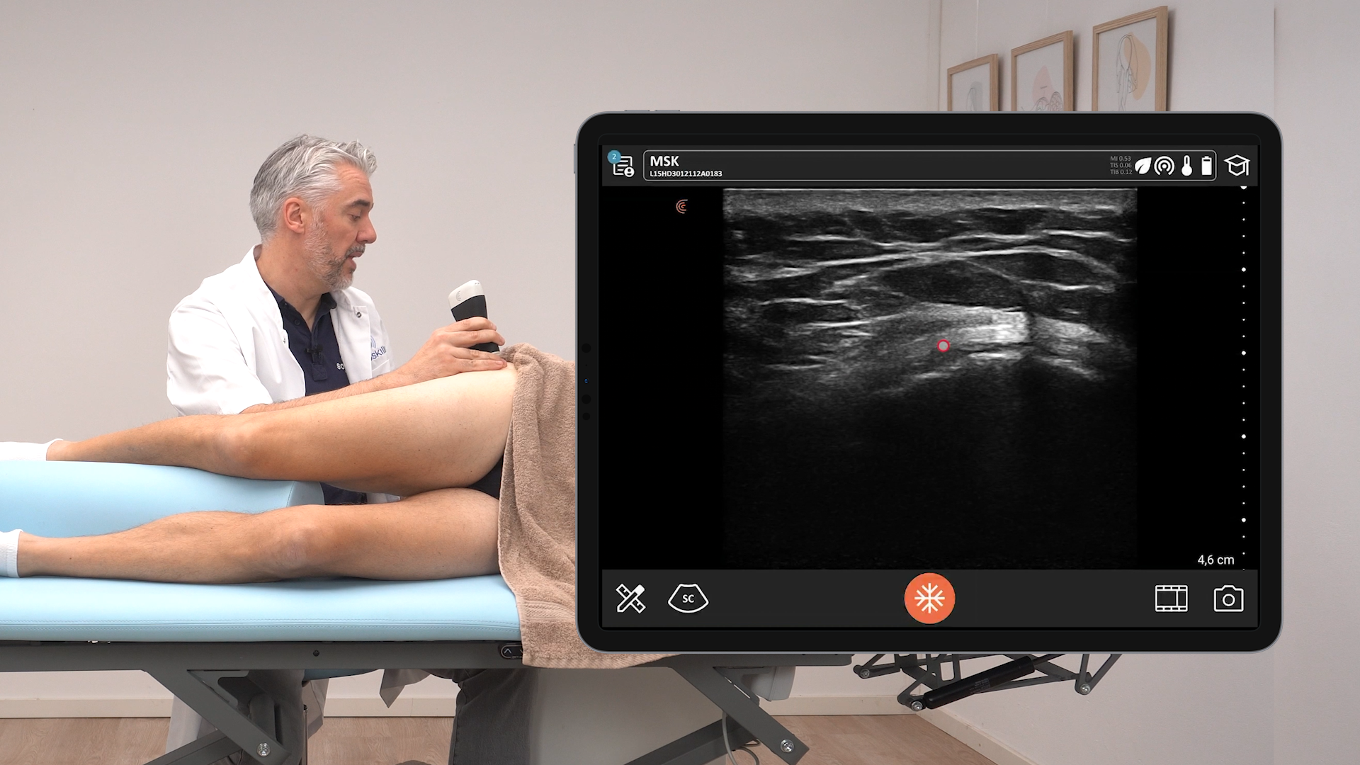
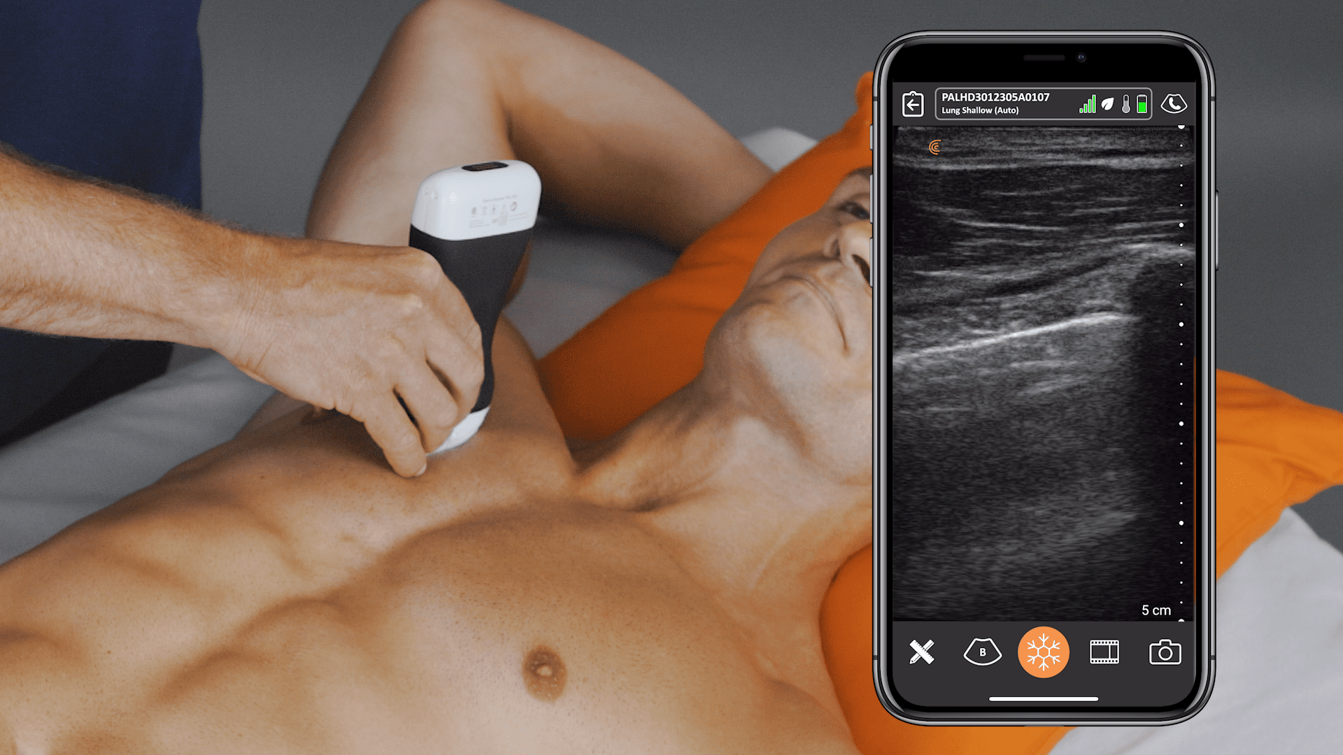
Our team worked with Dr. Dan Kim, a Vancouver emergency medicine physician, to record a quick lung exam tutorial using Clarius ultrasound.
Dr. Kim scanned a healthy model during the video. Please refer to the exam shown below from our cases page to see a clip of a lung with pneumonia captured with a Clarius scanner or visit our post on Clinical Utility and Technique for Lung Ultrasound in COVID-19 Cases for more detailed examples of lung ultrasound abnormalities.
Dr Shane Arishenkoff
Right sided lobar pneumonia acts as a window to visualize the heart from a right coronal view
Patient presented with cough, sputum, fever and hypotension. Large right sided lobar pneumonia identified. This is a coronal image taken from the right posterior axillary line. Initially the liver, diaphragm (right of the image) and hepatized lung (left of the image) can be seen. As the transducer is adjusted the beam is directed above the diaphragm and only the hepatized lung is seen. What is interesting about this image is the fact that the hepatized lung acts as a window to visualize the heart all the way from the right lateral chest.
Infection Control
During our time with Dr. Kim, we asked him about his thoughts on infection control concerns related to ultrasound use on COVID-19 patients and he had this to say:
With a lot of traditional cart-based units, it’s extremely difficult to clean all the surfaces and all the buttons and all the crevices. I think that there’s a role for handheld devices in this particular situation because they are much smaller than cart-based units and they are much easier to clean and disinfect compared to a cart-based unit.”
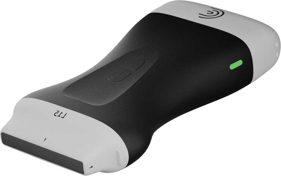
Small and wireless, Clarius scanners can be fully encased in a sterile cover. Clarius is IPX7 rated – it’s waterproof and fully immersible for disinfection. Review the long list of cleaners and disinfectants that are qualified for use with the Clarius Scanner.
Visit our Lung Scanning Resource page for more information.
