


Pulmonary embolism and other causes of right heart strain are often part of the differential diagnosis for patients presenting with chest pain or dyspnea. Relying solely on standard blood tests and radiography protocols can delay diagnosis, potentially leaving patients without timely, life- and function-saving treatment. A quick point-of-care ultrasound (POCUS) exam can make all the difference.
Dr. Tom Cook, an emergency physician and ultrasound educator, demonstrates the utility of POCUS to evaluate right ventricular function in patients with chest pain or dyspnea during a one-hour webinar that is available on-demand: Cardiac POCUS Part Two: Techniques for Assessing Right Ventricle Function.
Not ready to commit to a one-hour webinar? Scroll down for highlights of the discussion.
Understanding Right Heart Pressure and Dysfunction
Right ventricular dysfunction can be a critical finding in patients presenting with chest pain or dyspnea. The causes of elevated right heart pressure include:
- Left ventricular failure (most common chronic cause)
- Pulmonary embolism (most common acute cause)
- Isolated pulmonary hypertension
- Right ventricular infarction
- Pulmonary valve disease
Recognizing these potential aetiologies is crucial for accurate diagnosis and timely treatment.
Key POCUS Techniques for Right Ventricular Assessment
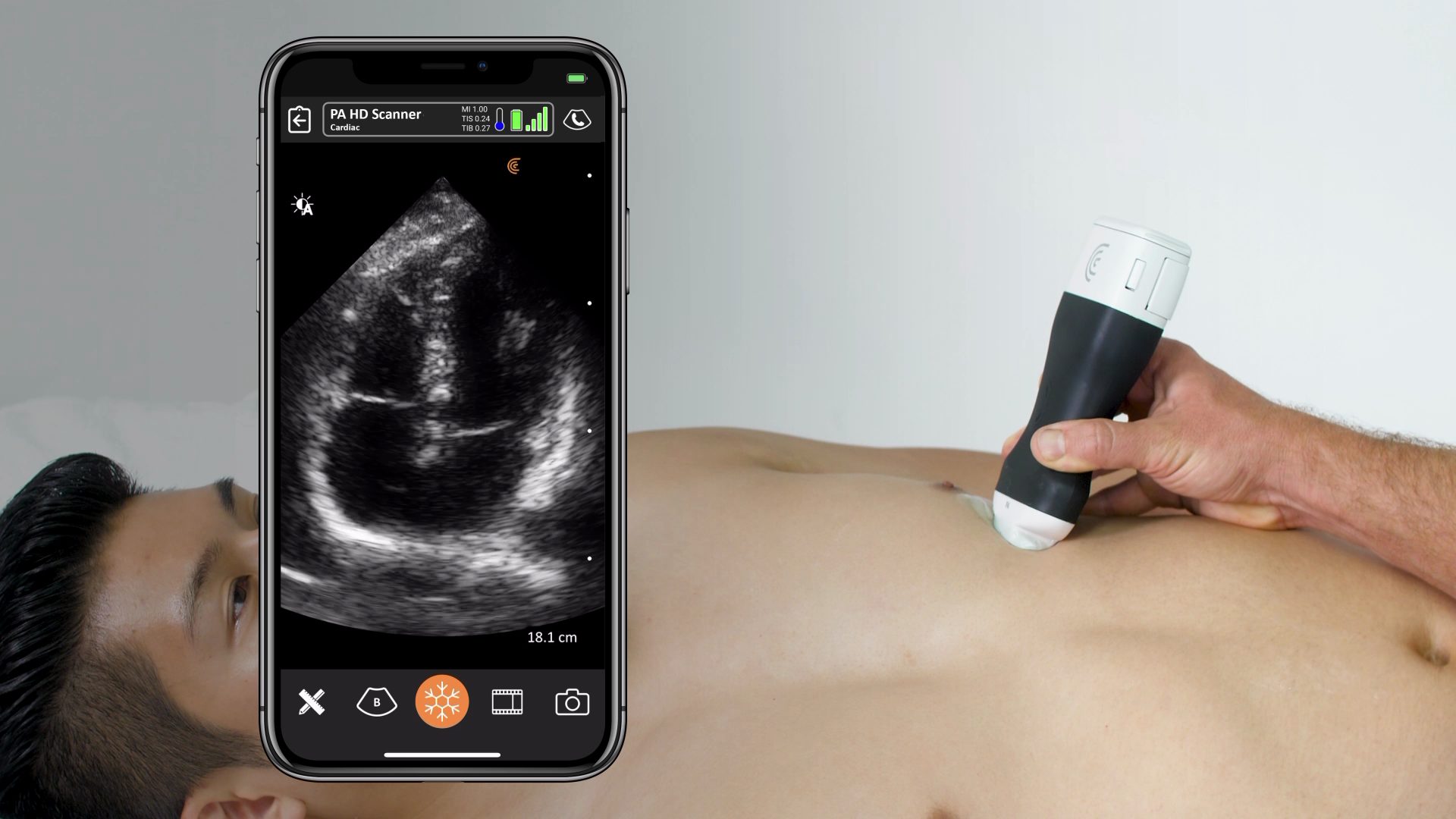
1. Apical Four Chamber View
This view provides an excellent comparison between the right and left ventricles. The right ventricle should be smaller than the left in a normal heart. An enlarged right ventricle compared to the left can indicate right heart strain.
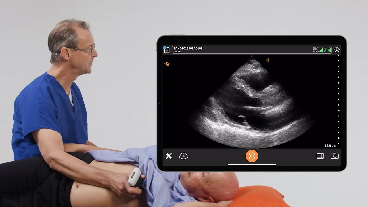
Watch this 2-minute video to see a demonstration of this advanced cardiac view that enables direct left and right ventricle function and comparison and mitral and tricuspid valve regurgitation analysis. Sonographer Shelly Guenther uses the Clarius PA HD3 high-definition wireless scanner for the demonstration.
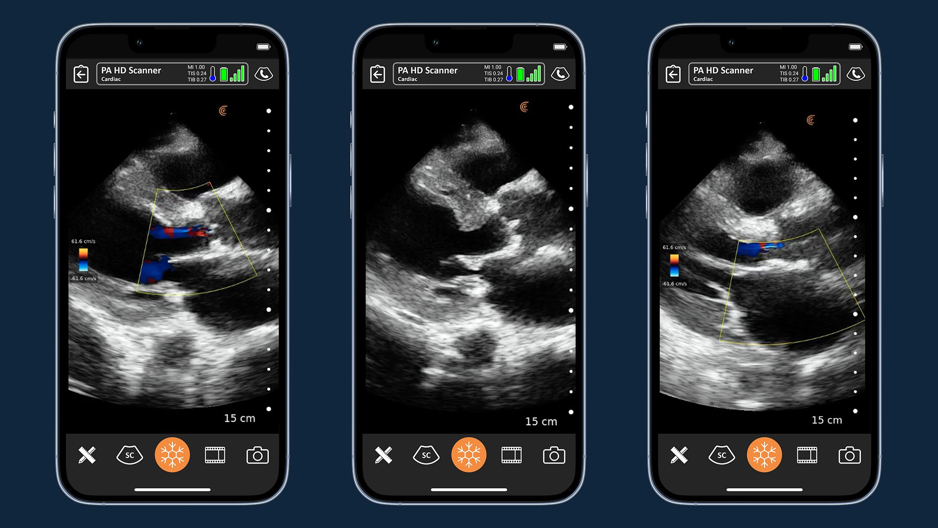
2. Color Doppler Assessment
Evaluating tricuspid regurgitation using color Doppler can provide valuable information:
- Flow convergence zone: The larger this area, the more pathological the regurgitation
- Vena contracta: The width of the regurgitant jet as it passes through the valve
- Jet area: The size and direction of the regurgitant jet in the right atrium
3. Coanda Effect
An eccentric jet that hugs the wall of the right atrium (Coanda effect) indicates a higher degree of pathology compared to a central jet.
4. Quantifying Pulmonary Artery Pressure
The color Doppler pattern of tricuspid regurgitation can be used to estimate pulmonary artery pressure. This non-invasive method can quickly provide crucial information about right heart function.
5. TAPSE (Tricuspid Annular Plane Systolic Excursion)
This measurement assesses the movement of the tricuspid annulus towards the apex during systole and is a reliable indicator of right ventricular function.
Case Study: Chronic Pulmonary Hypertension
Dr. Cook presents a case during the webinar where POCUS revealed:
- Enlarged right atrium and ventricle
- Moderate to severe tricuspid regurgitation
- Poor right ventricular wall contractility
- Small left atrium and ventricle
These findings, combined with the patient’s history, led to a diagnosis of chronic pulmonary hypertension.
Watch the full webinar to learn more about how integrating POCUS into clinical practice can significantly enhance patient care in emergency and critical care settings. Dr. Cook provides detailed insights to help you rapidly assess right ventricular function, quickly identify right heart dysfunction, estimate pulmonary pressures, and make timely, potentially life-saving diagnoses.
Clarius for Emergency Medicine: Affordable and Easy to Use Anywhere
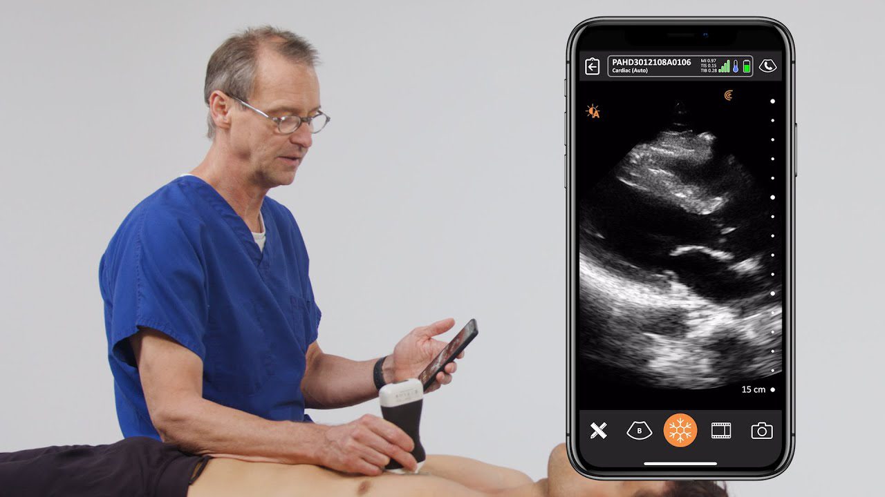
Dr. Cook primarily uses the Clarius PAL HD3. Wireless and app-based, Clarius ultrasound delivers fast imaging and sharp detail at the bedside. Visit our emergency medicine page to learn more about which scanner is suitable for your practice. Or book a virtual demo with a Clarius expert in your region.


