
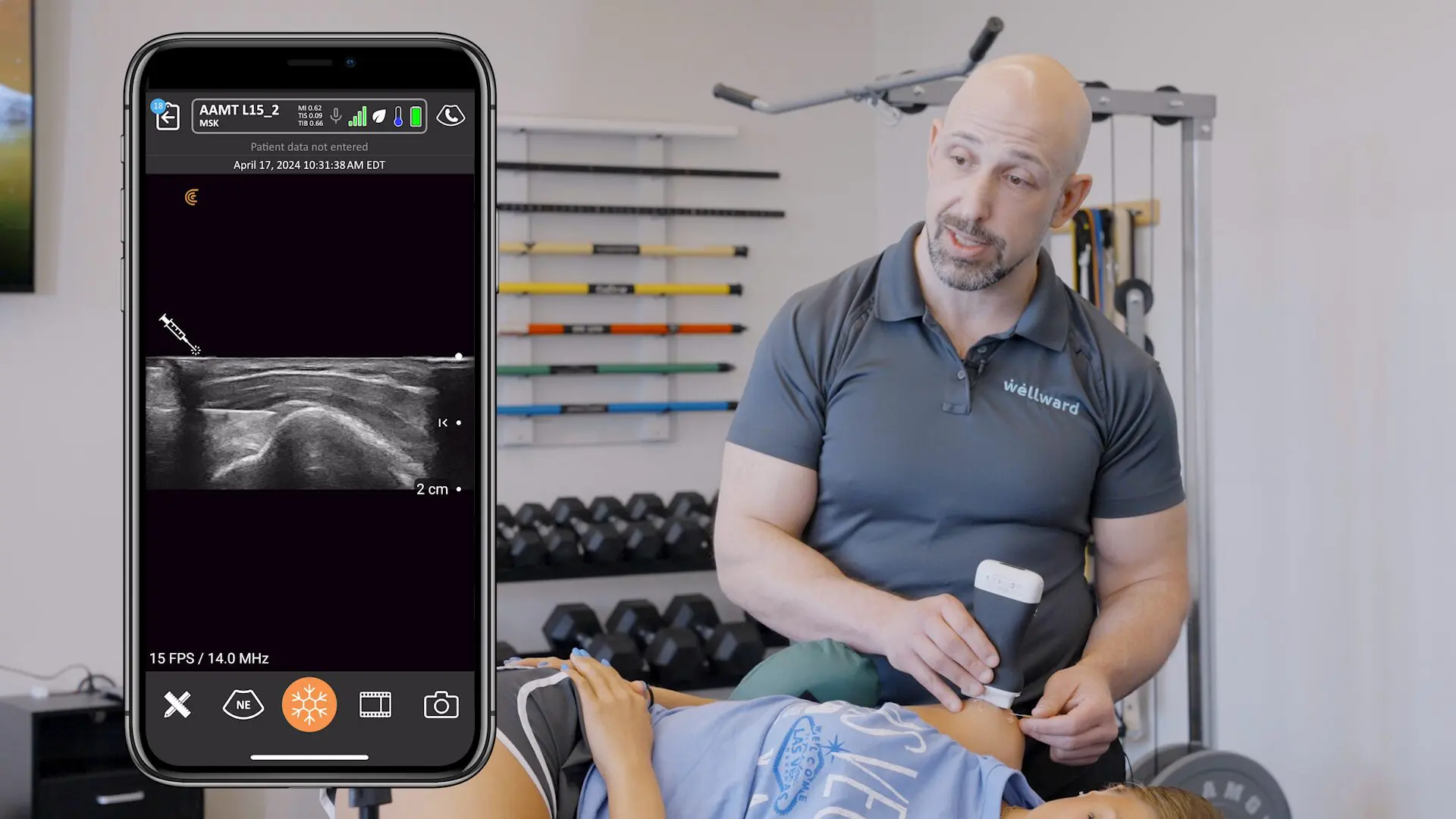
As ultrasound scanning is a new skill for many of our podiatry users, we’ve developed a series of educational videos and narrated examples of common pathology with Dr. Oron Frenkel an emergency physician who leads the Clarius Medical Advisory Board.
Scroll down to watch some examples below and visit Clarius Classroom to see the complete library.
Learn How to Scan the Heel with Clarius Ultrasound
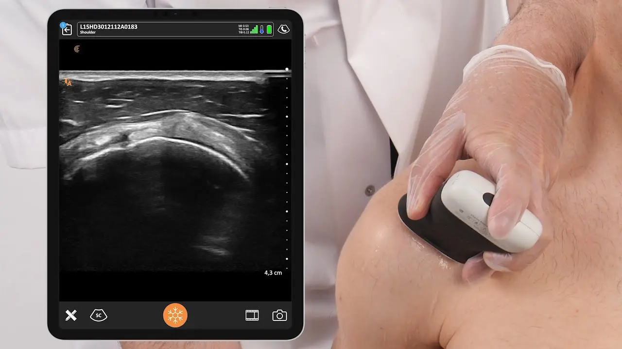
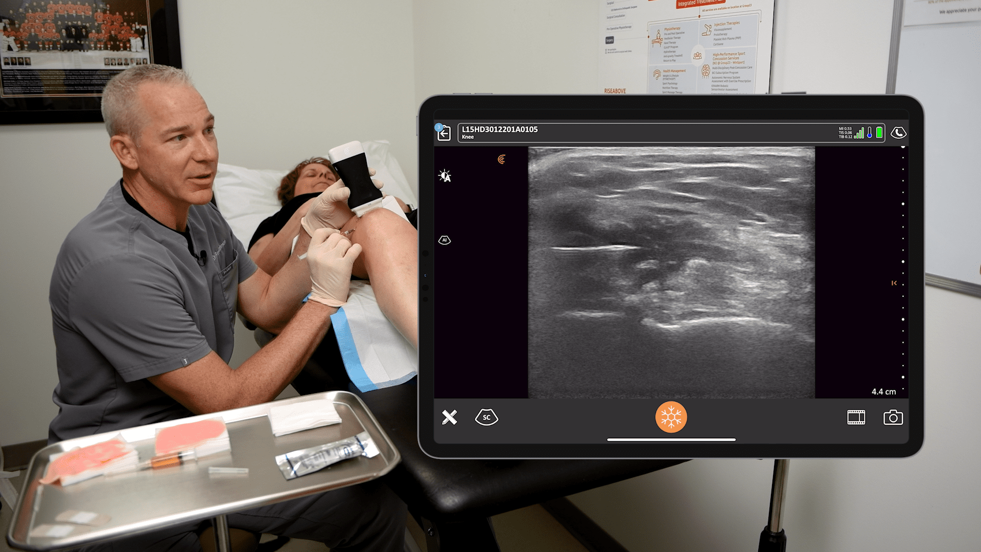
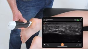
Dr. Frenkel demonstrates how to quickly scan a patient’s foot to identify signs of plantar fasciitis. He uses the plantar preset on the advanced MSK preset package that is available with Clarius HD linear ultrasound scanners. Watch the following video to see what a case of plantar fasciitis looks like with ultrasound.
See How to Identify a Case of Plantar Fasciitis Using Ultrasound
In the video above, you see an ultrasound scan of the thickened plantar fascia, which is best identified in the longitudinal axis from its insertion into the calcaneus. This is essential to diagnosing plantar fasciitis.
Learn How to Scan the Achilles Tendon
Palpation and patient history are often not enough to diagnose Achilles tendonitis. Watch this video to quickly and confidently scan the Achilles tendon to identify a rupture, tendinopathy, or tears of the gastrocnemius muscle. Scroll below to see videos of Achilles tendinopathy.
Identify Achilles Tendinopathy at Insertion
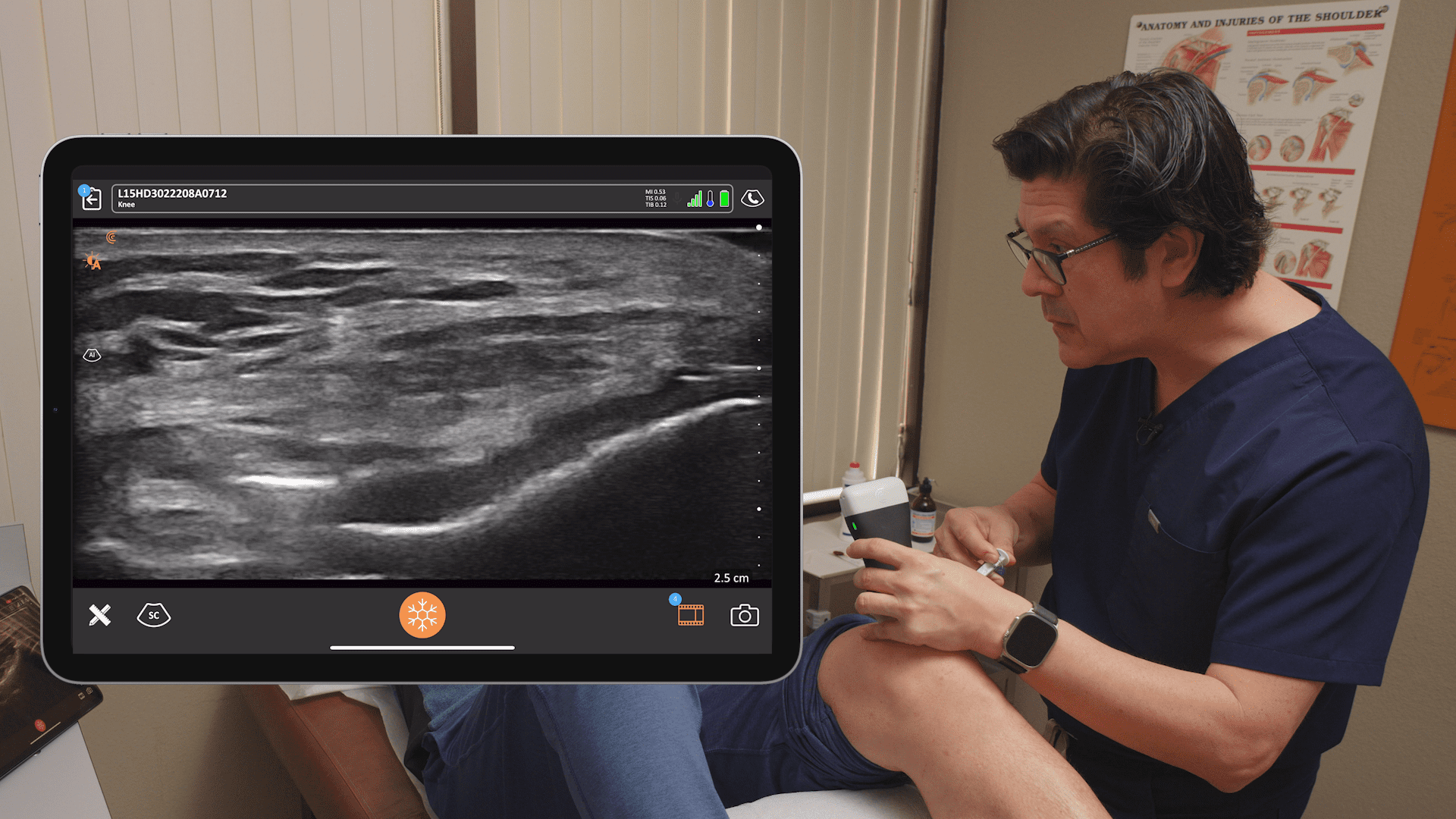
The video above depicts the long axis view of the Achilles tendon insertion to the calcaneus. You can identify multiple features suggestive of chronic tendinopathy, including enthesophyte, disorganized tendon fibers, calcification, and fluid surrounding the tendon.
Identify Achilles Tendinopathy at Midspan
The above video shows the short axis view of Achilles tendon proximal to insertion, which can identify heterogenous fibers, tendon edema, and partial or even complete disruption of the tendon fibers.
Identify Achilles Tendinopathy – Short Axis
In this video, an Achilles’ tendon suspicion of active inflammation is confirmed with increase signal using Power Doppler on the area of interest. A normal tendon should have little to no signal.
AI Automated Handheld Ultrasound for Podiatry
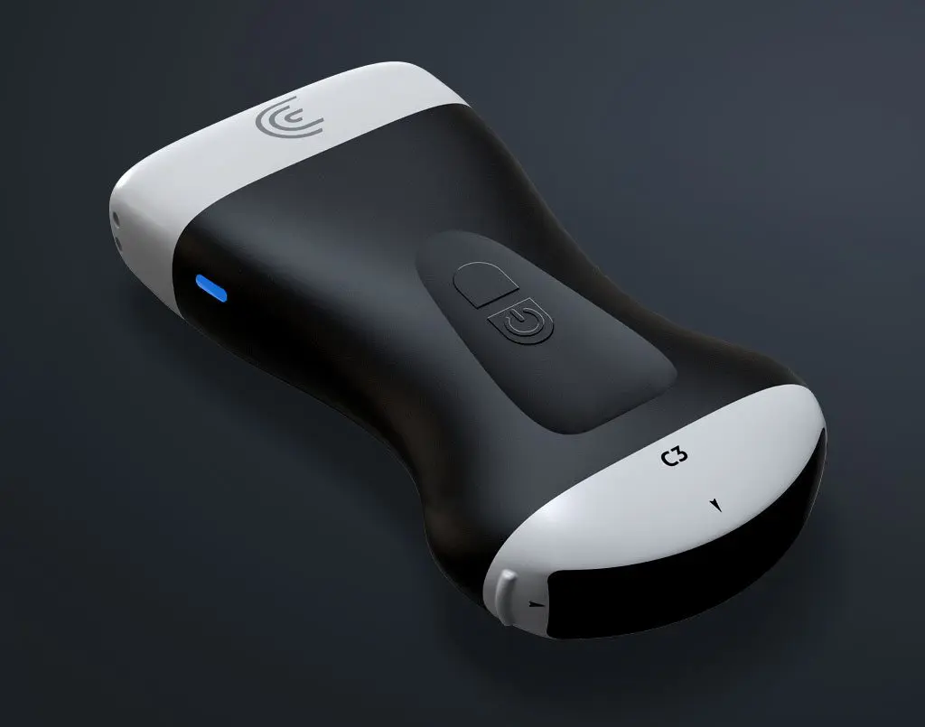
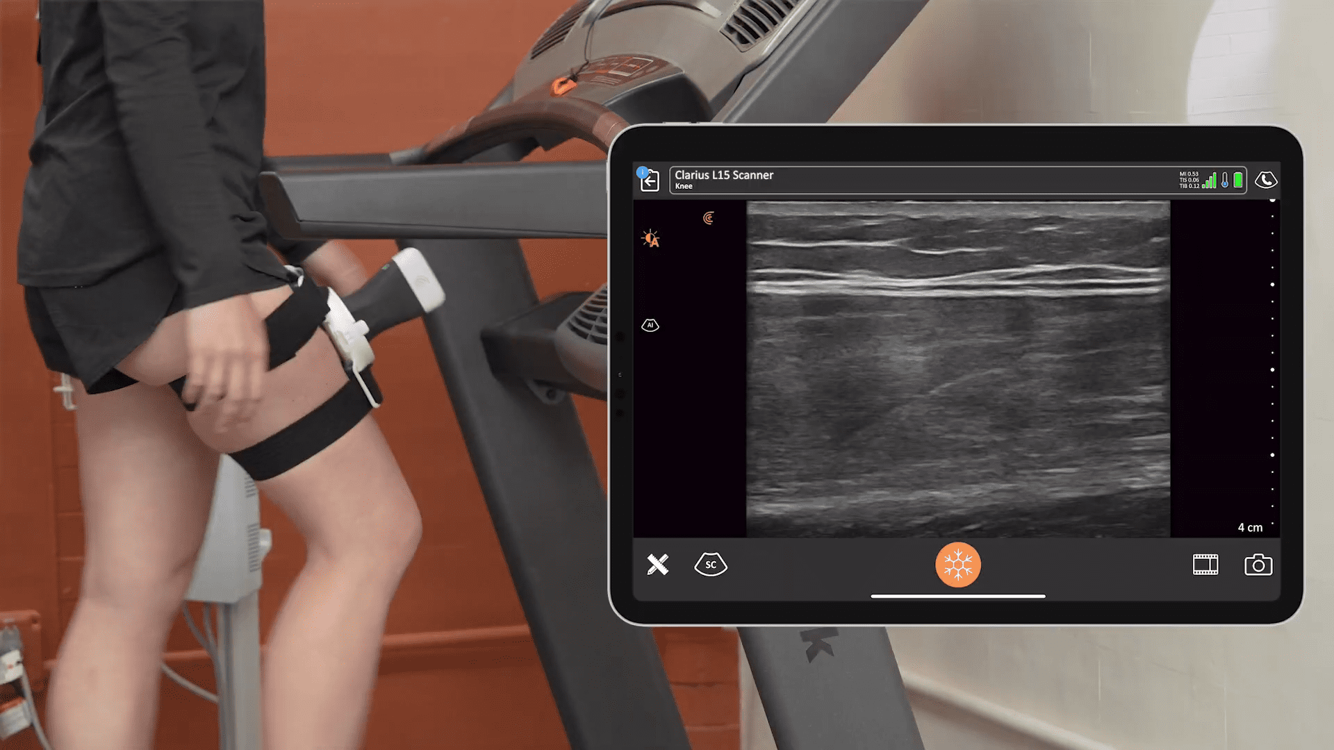
Getting detailed ultrasound images of musculoskeletal anatomy to make a confident diagnosis has never been easier or more affordable. Ideal for MSK anatomy down to 7 cm, the Clarius L15 HD high frequency ultrasound scanner is available with an optional Advanced MSK Package for diagnostic and interventional procedures. Each advanced Preset, including Foot/Ankle and Plantar, instantly optimizes the settings so you’ll get a clear, detailed image without adjusting complicated with knobs and buttons.
To discuss adding Clarius HD to your podiatry or MSK practice, contact us today or request an ultrasound demo. Or see come see live scanning at the Clarius booth at APMA 2021 this week!


