
Wireless ultrasound continues to advance the medical field, with guidance during medical procedures and interventions allowing for better visualization, accuracy, and outcomes. When it comes to injections into and around the Achilles tendon in particular, ultrasound guidance helps clinicians direct the needle toward the exact target.
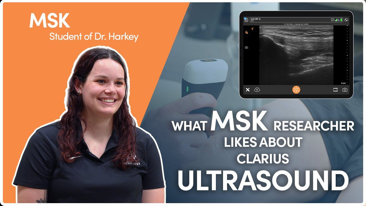
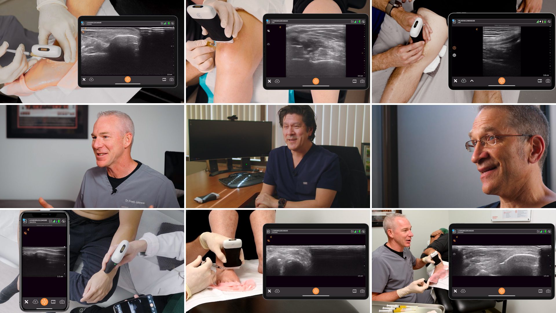
Clarius is excited to be working with Dr. Alan Hirahara for a webinar series on ultrasound-guided MSK injections. Dr. Hirahara began using ultrasound to guide his injections in 2009, noticing that his patients experienced significantly less pain after these ultrasound-guided procedures. Since then, he has developed new techniques to make injections more precise, easier, and less painful for the patients, which he shared with us during three webinars.
A recording of Dr. Hirahara’s latest webinar with Clarius, Ultrasound for Accurate MSK Injections and Interventions, Part 3: The Achilles Tendon, is now available to watch on-demand. Read the blog for highlights and video snippets from the webinar on how to use high-resolution ultrasound to best visualize needle placement in and around the Achilles tendon.
Choosing the Right Biologics
When it comes to treating tendon injuries, Dr. Hirahara explains the need to emphasize “how we can harness the body’s own regenerative abilities and healing properties.” This is possible with OrthoBiologics, which works with the body’s natural healing process. In his experience, a white cell inflammatory response resulting from leukocyte-rich platelet-rich plasma (PRP) increases inflammation, pain, and cell death while also creating a zone of weakness. As such, Dr. Hirahara recommends Leukocyte Poor PRP, in which platelets decrease inflammation, promote stem cell migration, reduce pain, inhibit bacterial growth, and create vascular channels.
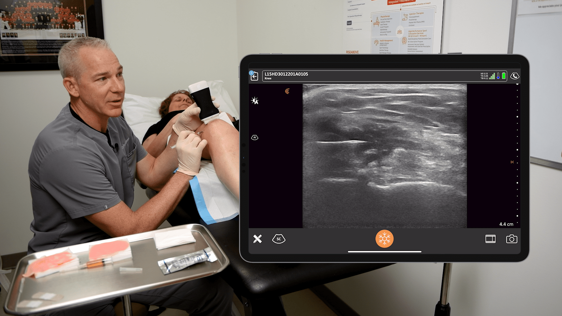
The benefit of PRP injections, compared to cortisone injections, is that the needle can go directly into the injured area, whereas cortisone injections carry the risk of rupturing the tendon. PRP injections target the injury more directly, especially with ultrasound guidance to ensure that OrthoBiologics goes exactly where it’s needed.
The Ultrasound Advantage
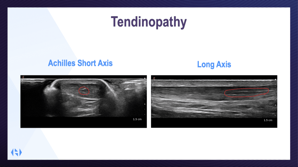
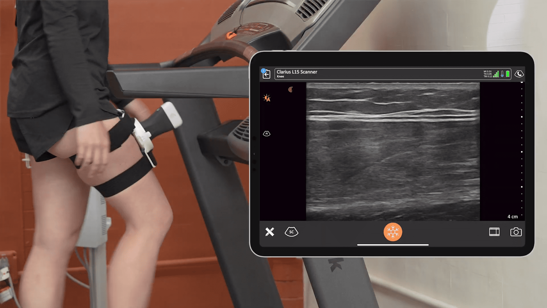
During the webinar, Dr. Hirahara showed some example ultrasound images taken on both the short and long axes, which are essential for getting the complete picture of the injury. He says that going through section by section is the best practice to ensure that the full extent of a tear is understood.
He emphasizes that having ultrasound imaging, which can see the subtle fibers of the tendon, is a critical and significant advantage that leads to better outcomes for patients.
Treating Achilles Tendinopathy
Traditionally, the technique for treating Achilles tendinopathy is in-plane long-axis injections. However, Dr. Hirahara quickly modified his technique because long-axis injections leave you inhibited by the leg, making it challenging to change the angle of approach and see what you’re doing.
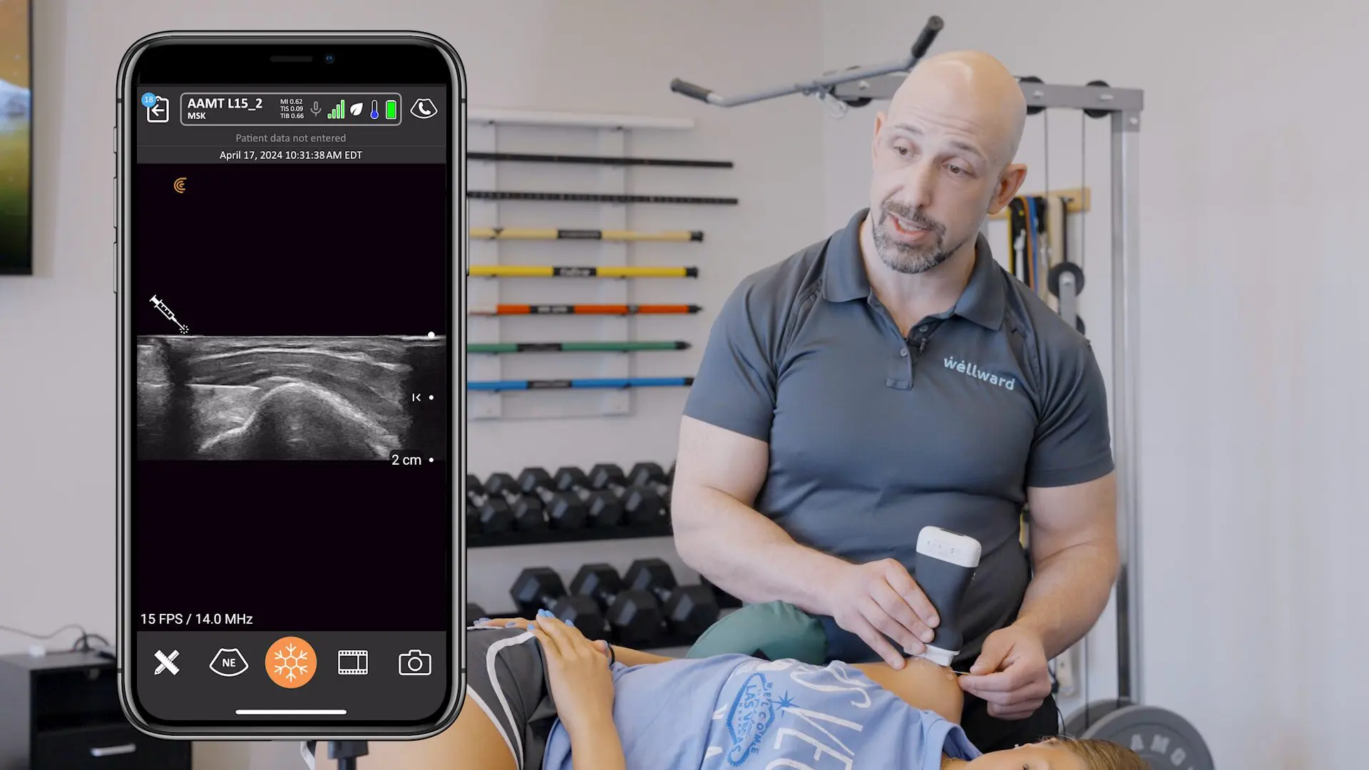
Instead, Dr. Hirahara does a short axis in-plane injection, which allows him to reach different parts of the tendon, including the peritendon area and sheath. He explained that he finds this technique to not only be easier, but it allows more flexibility regarding where the injection is going to go. Additionally, this technique allows him to access multiple sites of the tendon from a single insertion instead of having to poke the patient again and again.
Watch the Clarius Classroom video:
Aiding Achilles Tendon Rupture PARS Repair
PARS repair is a percutaneous method of passing sutures through an Achilles tendon, repairing the Achilles at the midsection or near the insertion point using a minimally invasive single incision.
As for PARS repair of Achilles tendon ruptures, one of the most common challenges is that by going in percutaneously, it is hard to tell whether or not the needle is actually passing through the tendon. This is because this procedure utilizes a jig, and if the jig is held too anterior or posterior, the entire procedure is essentially completed blind. When this occurs, it’s possible for the suture to miss the Achilles tendon entirely when passing the needles.
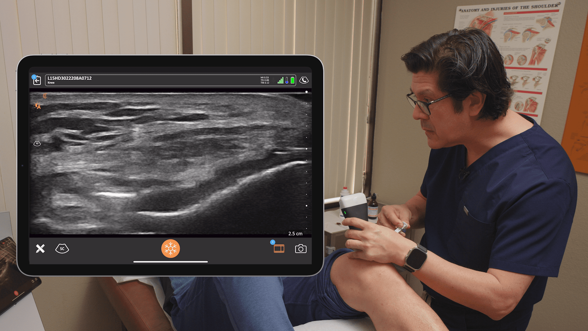
Dr. Hirahara uses his Clarius ultrasound to visualize the Achilles tendon and exactly where the needles pass through it. The use of ultrasound makes the procedure much simpler, and since the procedure is more straightforward, it can also be completed more efficiently.
Watch the video to see Dr. Hirahara demonstrate his ultrasound-guided technique:
Making the Most of Ultrasound Techniques
Dr. Hirahara recommends making minor adjustments to where you place your ultrasound scanner during the procedure. Those new to ultrasound may place the ultrasound and think they’re in the right spot, but tilting the scanner front and back or side to side allows you to optimize the image with ease.
Be mindful when scanning that what looks like a tear may just be anisotropy. Tilting your probe back and forth allows you to image more perpendicular to the tendon fibers, eliminating anisotropy and improving the overall image.
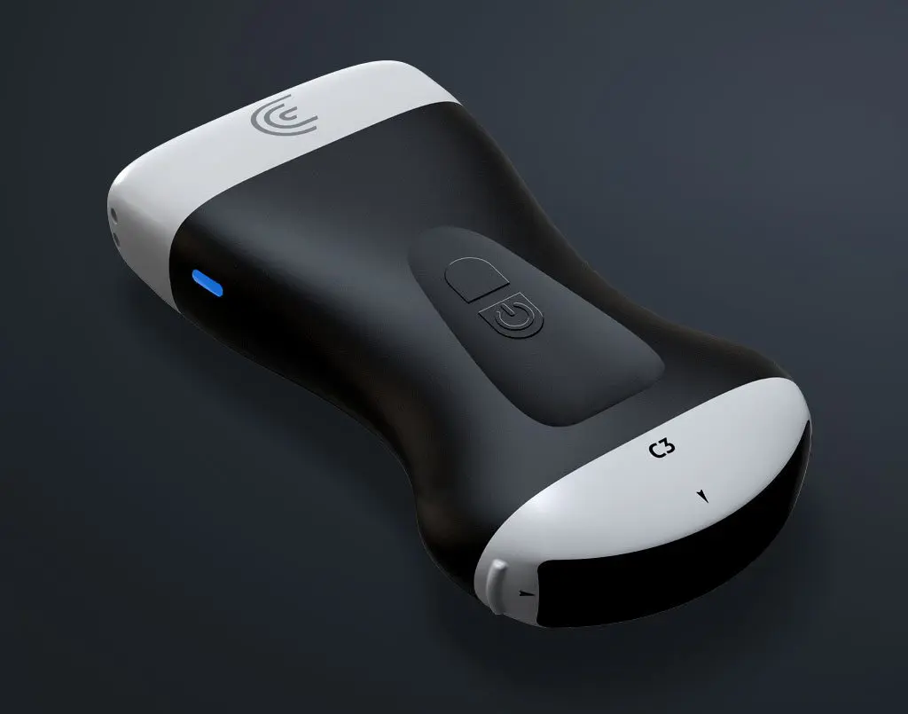
Eliminate Guesswork with Clarius HD3 Ultrasound for MSK Procedures
Clarius scanners offer image quality comparable to compact ultrasound systems for only a fraction of the cost, thanks to innovation and miniaturization. The Advanced MSK Package, included with membership, offers more flexibility for users who need additional customizations for MSK ultrasound.
Clarius MSK AI is a new advanced feature, currently only available in the US, that analyzes the tendon in real-time, places a color overlay that highlights the tendon, measures the thickest part, and labels it. Even Dr. Hirahara, with his many years of ultrasound experience, finds great value in the MSK AI’s ability to quickly note the thickest part of the tendon and increase reliability in the measurements.
To learn more about the high-frequency Clarius L15 HD3 wireless handheld ultrasound scanner Dr. Hirahara uses, contact us today or request an ultrasound demo. You can also see the difference high-definition imaging makes for orthopedic surgery by visiting our Orthopedic Specialty Page, where you can access additional Webinars, Testimonials, and Classroom Videos.


