1
00:00:00,110 --> 00:00:06,870
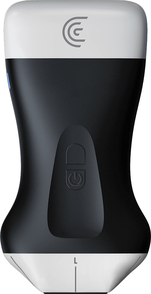
In this opportunity, we are going to talk about how to use the ultrasound machine to be able to perform controls for
2
00:00:06,910 --> 00:00:13,790
gastric balloon insertion. Gastric balloons, like any device, have an anatomy and
3
00:00:13,790 --> 00:00:19,709
have a shield, just like the breast implant, gluteal implants, and facial implants.
4
00:00:20,150 --> 00:00:26,390
This then generates an ultrasound pattern in its SHIELD or in its shields. Why is it important
5
00:00:26,390 --> 00:00:31,950
to perform an ultrasound control after a gastric balloon implant? Because we will be able to
6
00:00:31,950 --> 00:00:38,790
measure the volume of this implant, and we can see it exactly as it happens in the pleura.
7
00:00:38,830 --> 00:00:45,710
We can see the movement of the sinus inside the stomach. This makes us
8
00:00:45,710 --> 00:00:51,430
immediately see and diagnose that it is an implant that has adequate movement inside the
9
00:00:51,430 --> 00:00:58,389
stomach and what the final volume is. Next, we will see how we perform it.
10
00:00:58,669 --> 00:01:04,789
Important: I use this ultrasound machine for abdominal wall and abdomen patients, the
11
00:01:05,629 --> 00:01:11,000
HD three, which gives us the possibility to go from low frequency to high frequency.
12
00:01:12,440 --> 00:01:18,360
We are going to divide our abdominal wall into supraumbilical and infraumbilical, and we are going to
13
00:01:18,519 --> 00:01:25,480
use abdomen. In this way, we are going to look for the region at the
14
00:01:25,480 --> 00:01:30,640
subxiphoid level. We are going to look for our gastric image at the subxiphoid level.
15
00:01:33,320 --> 00:01:40,079
Always try to examine your patients standing up. Look then at how we see our
16
00:01:40,080 --> 00:01:46,960
spherical image, our wall, and the sphere. We are going to divide our screen and
17
00:01:46,960 --> 00:01:53,200
we are going to rotate our transducer clockwise to be able to see our
18
00:01:53,200 --> 00:01:58,160
implant volume in the stomach.
19
00:02:00,120 --> 00:02:07,000
What do we observe here? We obviously observe the walls of the implant. We are going to take
20
00:02:07,000 --> 00:02:12,840
the two measurements. The sagittal
21
00:02:14,679 --> 00:02:20,880
cut, and we are going to take the measurement in the axial
22
00:02:21,600 --> 00:02:28,360
cut. In this way, we are going to be able to have the result of what
23
00:02:28,360 --> 00:02:34,640
the volume of the gastric balloon implant is inside the stomach.
24
00:02:35,360 --> 00:02:42,039
Next, we are going to analyze our preperitoneal fat. There,
25
00:02:42,039 --> 00:02:48,479
I am going to change my setting and put it on abdominal wall to use the highest frequency
26
00:02:48,480 --> 00:02:55,360
of our HD three paddle. And at the midline level, at the linea alba level, what we are
27
00:02:55,360 --> 00:03:01,920
going to find in deep planes is the preperitoneal fat; this fat at the subxiphoid
28
00:03:01,920 --> 00:03:08,640
level we are going to measure in the postoperative period or the
29
00:03:08,920 --> 00:03:15,839
immediate insertion of the balloon. This is going to give us a prediction of cardiometabolic
30
00:03:15,840 --> 00:03:22,480
disease, and we can see how this measurement or thickness decreases as she
31
00:03:22,480 --> 00:03:29,089
loses weight. Remember that this preperitoneal fat with a metabolic
32
00:03:29,089 --> 00:03:35,690
concept is important to do a quantitative and qualitative follow-up. In this
33
00:03:35,690 --> 00:03:42,329
same way and with the linear and very high-frequency transducer, I return to the subxiphoid area and
34
00:03:42,330 --> 00:03:48,890
we are going to look for the implant wall here. Next, we can see
35
00:03:49,249 --> 00:03:56,249
what our implant and our shield look like. We see how there is movement during
36
00:03:56,250 --> 00:04:03,209
breathing and movement of the implant wall. This lets us clearly
37
00:04:03,210 --> 00:04:09,129
see and gives us peace of mind that the implant does not have a contracture and has an
38
00:04:09,130 --> 00:04:16,049
adequate displacement in the gastric fundus. This is the quick
39
00:04:16,049 --> 00:04:21,729
and reliable use of the ultrasound machine for any gastric balloon insertion procedure in
40
00:04:21,730 --> 00:04:23,649
metabolic patients.