












Clarius Classroom

T-Mode™ Shoulder for Physical Therapy Education

Dr. Christopher 'CJ' Miller

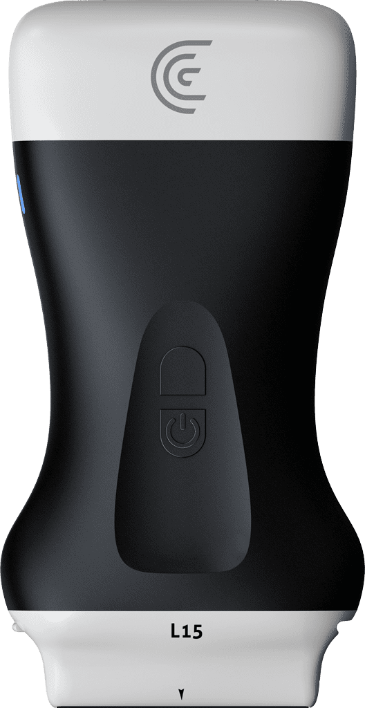
In this educational video, physical therapist C.J. Miller from Frontline Function demonstrates how to use the AI-driven T-Mode feature on the Clarius L15 scanner to evaluate the anterior shoulder.

Key Highlights

Palpation and Setup: The exam begins with the patient’s palm facing up while Miller palpates the anterior shoulder to locate the greater and lesser tubercles, which saves time before placing the scanner.

Bicep Groove Navigation: Placing the probe directly on the anterior shoulder reveals the bicep groove, positioning the lesser tubercle on the left side of the screen and the greater tubercle on the right.

T-Mode Activation: Once T-Mode is activated, the AI automatically highlights and labels anatomical structures, coloring the bicep tendon blue in its short axis and the overlying deltoid muscle orange.

Subscapularis Evaluation: By having the patient externally rotate their arm, the subscapularis tendon is brought into view in the long axis, with T-Mode dynamically highlighting its insertion onto the lesser tubercle.

Educational Value: Miller emphasizes that T-Mode is an excellent tool for educating both patients and fellow clinicians, as it instantly identifies unknown structures and allows the user to easily toggle the color overlay on and off to view the true ultrasound image.
Specialties: MSK, Physiotherapy
Applications: MSK, Shoulder

My name is C.J. Miller, and I'm a physical therapist with Frontline Function. Part of my role is to see patients in the orthopedic and primary care settings. But a lot of my role is to educate physical therapists on how they can integrate musculoskeletal ultrasound into their practice. Today, I'm going to use the Clarice L 15 scanner to demonstrate T mode, which is an excellent tool for education purposes.
The first thing I'll do is put our patient with his palm up and I can palpate the anterior shoulder, finding the greater and lesser tubercles. This just saves me time searching. Once I can find that, we'll go directly onto the anterior shoulder and we'll be able to find our bicep groove with the lesser tubercle on the left side of the screen and the greater tubercle on the right side of the screen.
Now, if we activate T mode, this is going to fill in everything we're looking at really well. So the tendon in the middle of the screen on the left highlighted in blue on the right labeled tendon is a short axis of our bicep tendon. I can scan up and down and it will monitor where that tendon goes
01:07
–
02:09
Speaker 1
above. That muscle typically shows up as dark on the right side, labeled deltoid on the left, labeled orange. If I want to see the true image, I can turn off the coloring on the left side. Furthermore, if I want to evaluate other tendons with this T mode, I can have him externally rotate and bring our sub scap tendon in the view, and our T mode will highlight it for us so it can demonstrate where that sub scap tendon in long axis is running and inserting onto the lesser tubercle.
I can move him back to our view here, and our bicep tendon comes into view in short axis there as well. The T mode is a really nice educational tool because if you're searching for things and you're unsure what structure is what, this uses AI to automatically highlight the structures that are on your screen.
So that was a demonstration of T mode using the Clarice L 15, which is a great tool for educating both your patients and if you're educating your fellow clinicians.

Products Used

More Classes

Request a Quote

With 8 scanners available, we offer a wireless ultrasound solution tailored to your needs. Request a quote to discover which scanner can deliver the best ultrasound imaging for your practice.

⚠️ Clarius ultrasound is for medical professionals only.

By providing my email, I consent to receive Clarius webinar invitations, case studies, whitepapers, and more, and I consent to the Clarius Privacy Policy. I can unsubscribe anytime.