
An abdominal FAST exam provides real-time information about free fluid in the abdomen, pericardial and pleural spaces, which are often missed or only suspected through a physical exam, basic blood and urine testing, and even radiography. Ultrasound is now commonly used cage side at the point of care. It’s low impact, rapid, safe, radiation free, requires no shaving, and only minimal patient restraint – all advantages for your critically ill patients.
Although FAST exams started in emergency rooms, veterinarians are now using the exam for animals where anaphylaxis, pericardial fusions, pleural fusions, non-traumatic hemoabdomens and peritonitis is suspected. The abdominal FAST has been shown to help screen for anaphylaxis by looking at the halo effect on the gallbladder. It is used to look at cardiac function by looking at the caudal vena cava caver size and assessing volume status. It’s also used for looking at urinary bladder integrity.
With an Abdominal FAST exam, we can detect as little as two to three mls per kilo of free fluid. That’s something that an x-ray just cannot achieve,” says Dr. Camilla Edwards, an experienced veterinarian and ultrasound educator based in the United Kingdom. “Ultrasound detects a small amount of free fluid in the peritoneal, the retro-peritoneal, pleural, and pericardial spaces. We can also predict the degree of anemia in traumatic hemoabdomens in particular, and we can serially monitor for occult hemorrhage post intervention, such as biopsies or surgeries.”
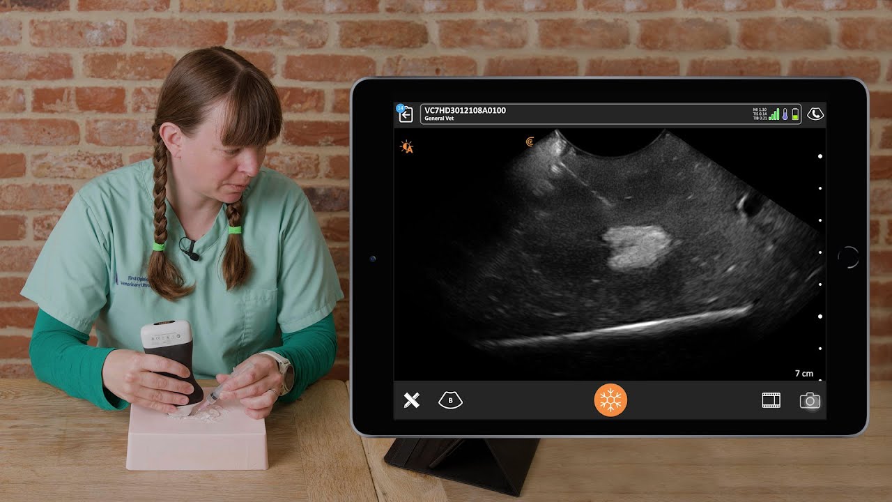
Dr. Camilla’s Abdominal FAST Video Demonstration
Watch this video for a step-by-step demonstration of an abdominal FAST exam performed on Dr. Camilla’s dog Pippa.
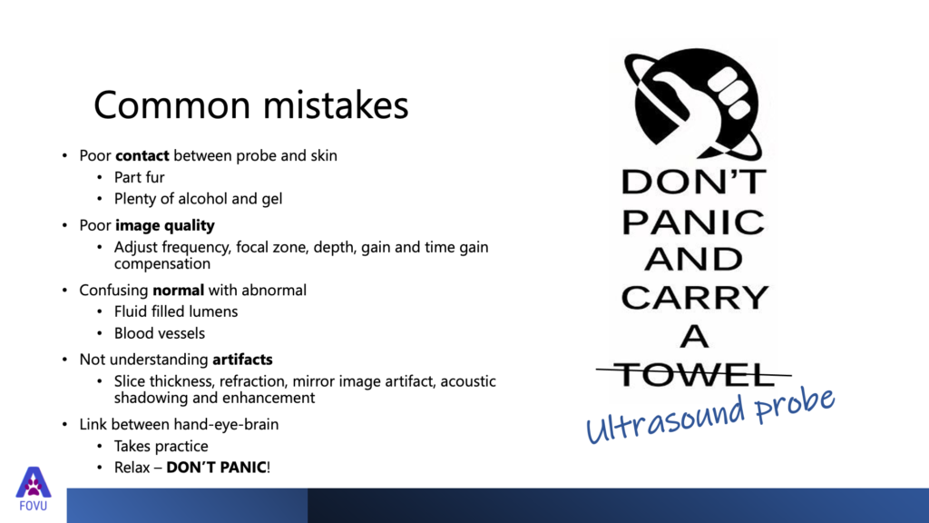
Common Mistakes with a Small Animal Abdominal FAST Exam
In a recent webinar presented by Clarius about abdominal FAST scanning techniques, Dr. Camilla shared the following common ultrasound scanning mistakes
1. Poor contact between the probe and the skin.
“Make sure you’ve parted the fur, because fur will trap air and air is the enemy of ultrasound and you won’t be able to see through it. Apply plenty of alcohol and a load of gel, and then you’ll get a good image. You can also just use alcohol to wet down the fur, and that is often enough.”
2. Poor imaging
“It’s important to learn how to adjust your frequency, focal zone, depth, gain, and time/gain compensation. It can make a real difference to your image and affects your diagnosis. The most important adjustment is depth. You’ll get a much better image of what you want to see if you adjust to the right depth.”
3. Confusing normal with abnormal
“I think lots of vets new to ultrasound are really worried about doing this. The things that you might confuse as abnormal but are actually normal might be fluid-filled lumens, such as the urinary bladder or the gall bladder, and blood vessels, which are long, thin tubes or round circles when they’re cut and transverse. Learn to recognize what common structures are supposed to look like.”
4. Not understanding artifacts
“There are a lot of artifacts that we see that give us a lot of information on ultrasound. It’s worth getting to know a bit more about those. For example, slice thickness, artifact refraction, mirror image artifact, acoustic shadowing, and enhancement. Those are really key artifacts to understand. If you can see your anechoic black fluid, that’s the important thing to watch out for first.”
5. The link between hand-eye brain coordination
“Learning to use ultrasound takes practice. Don’t worry and don’t be scared to try it. Pick up the probe and try. The more you try, the better you’ll get at it. My advice to new users is to relax, don’t panic, and carry an ultrasound probe.”
Typical Cases Seen in Veterinary Practice
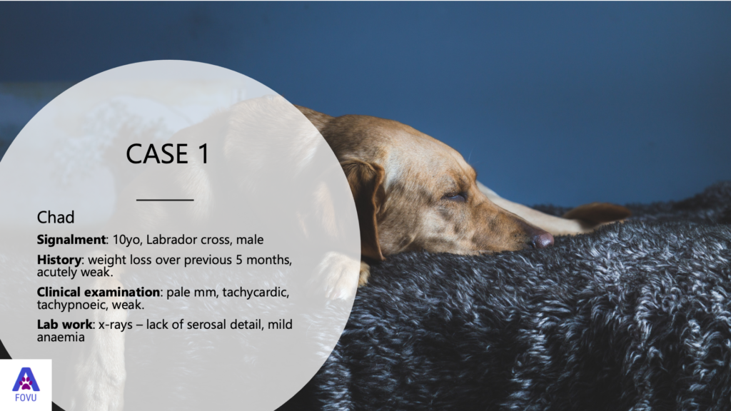
Abdominal FAST Exam on a Male Labrador
“Chad is a 10-year-old male Labrador cross. He’d been losing some weight over the previous five months, but he presented acutely weak, almost collapsed. It’s a fairly typical thing that we’ll see in the emergency shift in the evening, that the dog seems just very weak suddenly. When my colleague looked at the dog, he had pale mucous membranes, it was tachycardic, tachypnoeic, and extremely weak. The x-rays showed a lack of serosal detail, so that’s giving us a clue that there might be free fluid. Mild anemia was evident on blood work as well.

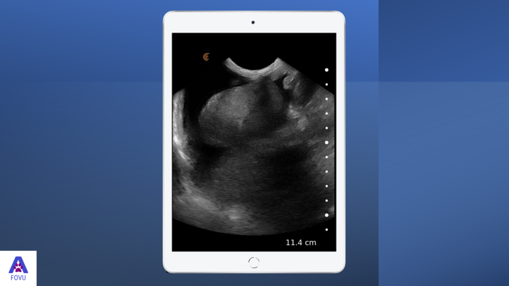
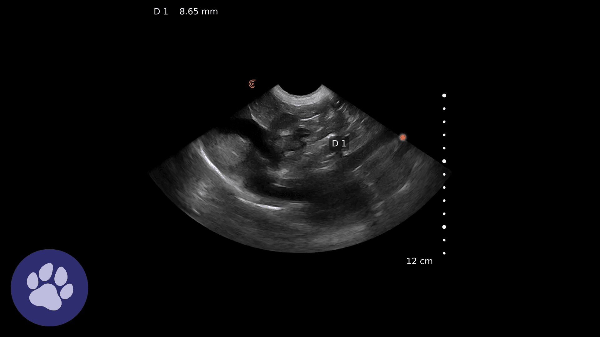
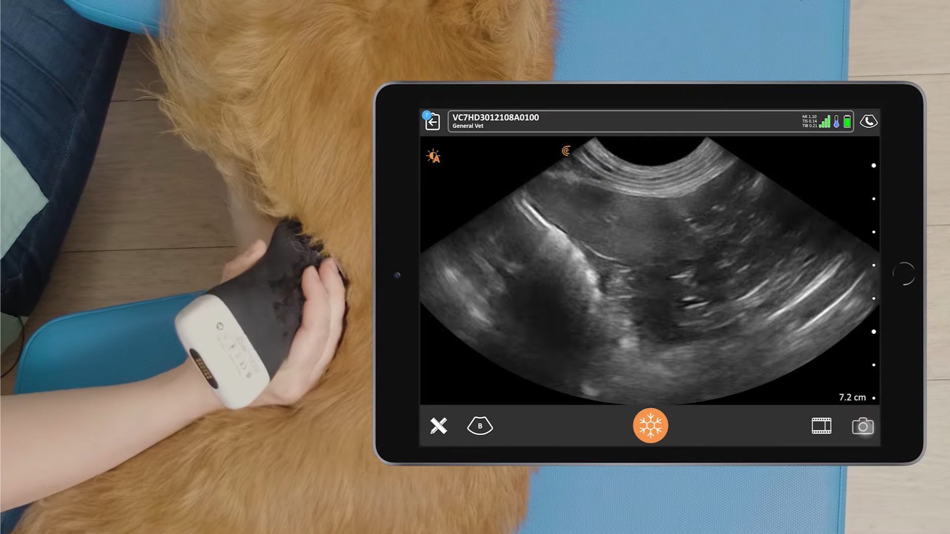
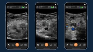
In these ultrasound images we can see some spleen and we’ve got some intestines. We’ve got the spleno-intestinal-umbilical view and this is quite a normal looking spleen. We’ve got a loop of intestine and all around it, we’ve got black fluid forming triangles, and that’s free fluid. We know it’s free fluid because it’s forming a triangular shape instead of a nice circle, which would mean it was a lumen.
To confirm what we see is free fluid, we performed an abdominocentesis. We used ultrasound to easily find the fluid pocket and avoid poking the needle in a loop of intestine. While this procedure has been done blind for a long time in veterinary medicine, even with a little training, it’s safer to see what you’re doing.
The abdominocentesis confirmed a hemoabdomen. Since there was no trauma history, this likely means we’ve got a splenic mass. With the concern around malignancy with hemangiosarcomas and the age of the animal, the owners opted to euthanize at this stage.”
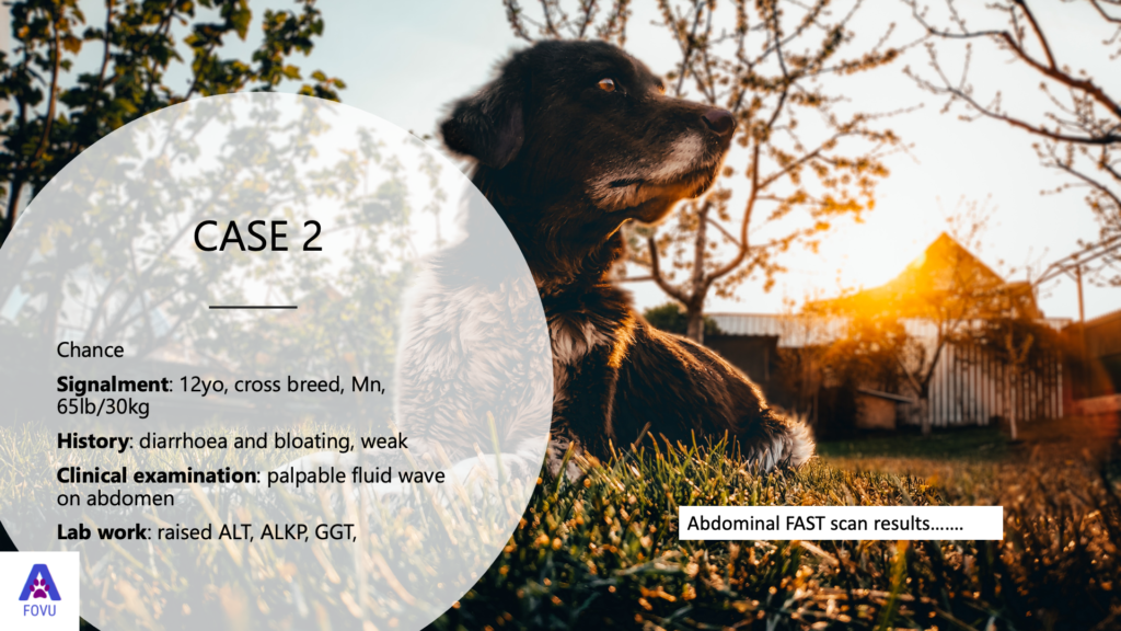
Abdominal FAST Exam on a Crossbreed Male
“Chance is a bit more of a chronic case. He’s a 12-year-old crossbreed, male, neutered, weighing in at 30 kilos or 65 pounds. He has a history of diarrhea and bloating. On clinical examination, my colleague found palpable fluid wave on the abdomen, and the lab work showed a raised ALT, ALKP, and a raised GGT. Let’s have a look at the abdominal FAST scan results.
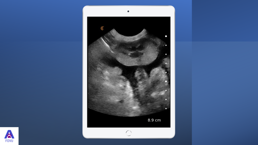
Diaphragmatico-hepatic view
We’ve got the diaphragm and the liver and we can see that there’s an enormous amount of fluid.
Spleno-renal view
The spleen is not quite in the image, but we’ve got the left kidney there. Then we’ve got mesentery and intestines down here, and we’ve got free fluid in between the kidney and the mesentery.
Cysto-colic view
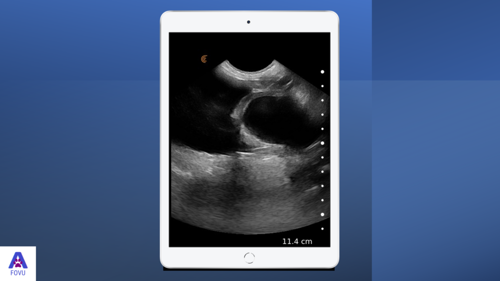
In this view we see the bladder. This shows you how round the bladder can be. When it’s starting to get equal pressure from inside and out, then it becomes quite round. We can also tell that since we’ve got a bladder with fluid, then it’s very unlikely to be ruptured as it will deflate without the fluid.
Intestines with mesentery in between
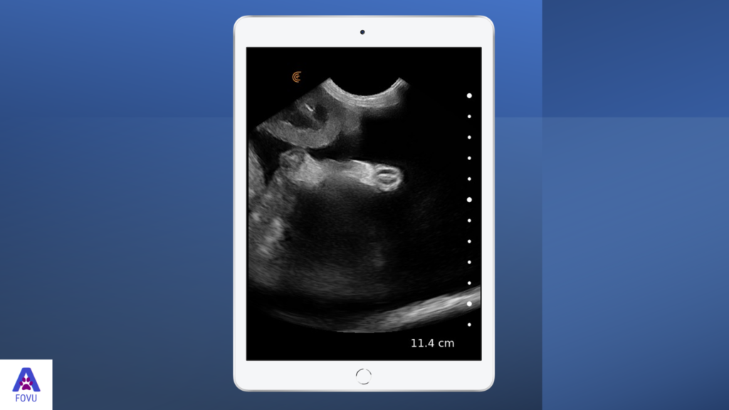
I often get asked about images like these in my courses because students are concerned about what these things are. This is intestines with mesentery in between. The black area is free fluid. We see enormous amounts of free fluid here. But these are normal looking intestines. It just looks very different when it’s highlighted by free fluid.
In this case, the abdominocentesis revealed clear yellow fluid. The patient was referred, and multifocal non-surgical tumors were found in the liver, and coincidentally also, choleolithiasis. The owners opted for palliative management, so the dog started on Gabapentin and diuretics and twice weekly abdominal drainage to take the pressure off the chest. The dog is still going six months later.”
Getting in Touch with Dr. Camilla
Dr. Camilla invites you to get in touch with questions via [email protected]. Or you can visit her website. You’re also invited to watch Dr. Camilla’s review of the Clarius C7V HD for small animal veterinary medicine.
Dr. Camilla runs online courses and teaches in practice. She also runs a Facebook community with discussions about veterinary ultrasound.
More Veterinary Ultrasound Education
We invite you to watch the full webinar with Dr. Camilla: Practical Small Animal Ultrasound AFAST Scanning Techniques. You may access more of Dr. Camilla’s tutorials on Clarius Classroom.
Interested in Affordable Ultrasound for Your Veterinary Practice? To learn about how easy and affordable it is to add Clarius handheld ultrasound to your veterinary practice, visit our Clarius veterinary ultrasound page for a video demonstration and product details. Or contact us today to discuss which scanner is right for your veterinarian practice.

