Hi, I'm doctor Tatiana Verloc. I'm an emergency physician and founder of. Hello, sono. We help launch compliance and high quality focus programs. Today, I'll walk you through how to use focus to evaluate patients with suspected congestive heart failure. I'd like to start with the cardiac scan first.
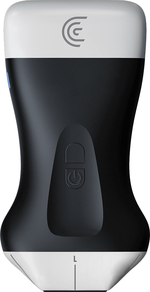
Starting with personal long axis view. I am using a multi-purpose Pal scanner. You can also be using a phased array scanner to get a good look at the heart.
In this case, note that my screen marker is on the right of the screen, so I will line up my probe marker to be facing patient's right shoulder.
And I'm starting at around third to fourth intercostal space in a personal region to get my first view.
Here it is. So in this view I decrease the depth a little bit. The biggest chamber here is the left ventricle on the left side of the screen. We are seeing the aortic outflow, aortic valve, left atrium and the mitral valve, and also our vault at the bottom of the screen. The circular structure is the descending aorta.
The first thing I look at is actually the movement of the anterior mitral valve leaflet. So if it's touching the intra particular septum it's getting very close to it. I know that the ejection fraction is good in this patient.
So let's just watch it a little bit more I am increasing the gain here. Here you go. I am then looking at the actual myocardium. And typically at that introvert articular septum and the bottom of a screen to here we want to see the myocardium thickening.
Another great tool you can use is EFI here which will do an auto ejection fraction.
Here you go. So it's showing a real time ejection fraction.
Our next view is the personal short axis view. I typically use two hands to turn my probe and face the probe marker to its patient's left shoulder. Here we should get a nice concentric view of the left ventricle with a papillary muscles visible. And you're looking for myocardial thickening and very symmetric squeeze of the left ventricle with around 50% change in the diameter of the left ventricle between Sicily and diastole.
For normal EF, we are also visualizing the right ventricle and the left side of the screen here.
Our next view is apical for chamber view and for that view, point the probe marker towards 1 to 3:00 and place the probe at patient's PMI. So we'll try to identify it here.
So around here
I'm going to increase the depth a little bit
and increase the gain.
So this is our four chamber view. This view can be used as well to look at that mitral valve leaflet. How close it's getting to the particular septum.
It can also be used to evaluate for diastolic function. So the base of the heart where tricuspid annulus is and the mitral valve analysts are those those are called the base of the heart. And you're looking for up and down movement. If there is a lot of movement there, that means that the heart is able to relax and fill.
So that correlates to a good diastolic function and is a quick way to tell without doing any complicated measurements. Our last view is the subcostal says I forward view. I will have the probe marker facing around 6:00, and I am using an overhand grip here because we need to have quite a low angle to get the heart into view.
I'm in this sub cipher area.
And ideally we want to be scanning through the liver.
I'm just going to increase my depth here.
So we've seen the heart and I increase the gain.
Here we go. This is a great view to look for pericardial effusion especially if it's at all on your differential. To improve this view, you can have the patient take a deep breath and hold. So can you try doing that. Just say breath in and just hold
Here we go. And after you obtain this view, identify where your rate HMS. Just right over here. Center it on the screen and rotate your probe 90 degrees to get the IVC entering the right atrium. And before doing that, I'm actually going to switch to the abdominal setting here.
Here we have a view of the IVC where it's entering the right atrium. And to assess the collapse ability of IVC, you want to be looking at an area about two centimeters distal to the right atrium, or just past the hepatic vein drainage.
If you are seeing the IVC being highly collapsible, meaning more than 50% of the walls are completely touching, that means that the tank is empty and the patient is not fluid overloaded. In fact, they are fluid down. If you are seeing that IVC being distended and there is very minimal variability with respirations, with respiration, that tells us that the tank is full and this is more likely, it's more likely that this patient is fluid overloaded.
Our final view is the view of the lungs. And I will demonstrate on the left side of our patient how to get the long views. First I'm going to change our setting to the long view.
And in this case I'm going to start with the left apex of the lung. And this is our second intercostal space microvascular lined with in the sagittal plane with a probe marker towards patient's head. And what I'm looking here for is first of all pleural sliding. I'm decreasing my depth to show that to you.
Also going to decrease the gain a bit. Actually press auto gain.
So here we clearly see that plural sliding. Telling us there is no pneumothorax. Another thing we are looking for are these horizontal lines
that are equidistant from each other and go down the long field. This is a reverberation artifact that presents that is present in a normal lung that's air filled. So in the case of CHF, instead of those air lines, you would be seeing vertical lines that are called B lines. And they emanate from the pleura and go down the screen.
And the more of them you see, the more of a disease burden there is. And you would see them bilaterally. Our next view is that posterior axillary view. You can also have the patient set up to visualize the posterior fields better. Here I'm in the coronal plane the probe marker up. And I'm looking for the same things.
I'm looking for that plural sighting and the aligns which we are seeing two zones here, and I'm moving my probe around trying to catch more areas. And our final view here is the long base.
I am still in the coronal plane. So here we got the spleen. If I move a little bit distal I'll visualize the kidney. However I do want to see that diaphragm right. That's that cost of retrieval angle that we also look at on a chest X-ray. So here's a clear view of the diaphragm and the spleen reflecting on the other side.
This is called mirror image artifact. And that's normal. When the lung is air filled the lung is on the left side of the screen. Another thing to notice here is the spine at the bottom of the screen here. And it ends at the diaphragm. That only happens when the lung is air filled. If there is fluid or consolidation there, the spine will continue.
And this is known as spine sign. So if there were a pleural effusion here, you would be seeing anechoic black fluid and the thorax and the continuation of the spine. So putting all of this together with a long ultrasound, you are looking for those B lines and pleural pleural effusion. With the IVC you're looking for local stability distended IVC.
And then finally with a cardiac ultrasound you're evaluating for decreased ejection fraction. Putting all of those together. If all of these findings are positive, your patient is likely having CHF exacerbation and needs diuresis. Thank you for watching. Please check out hello sono com if you'd like any help when launching a high quality and compliant podcast program, and do check out the rest of these high impact quick tutorials.