
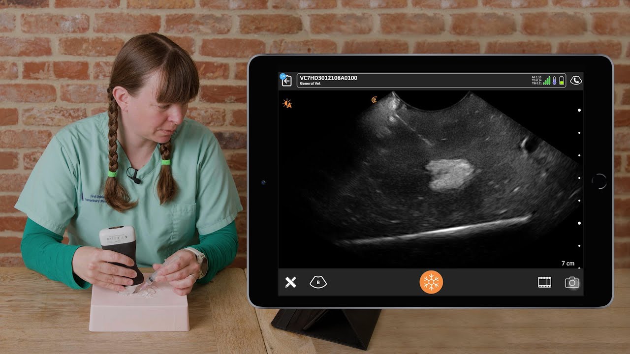
This week we’re adding more practical tips from Dr. Camilla Edwards, DVIM to our library of point-of-care ultrasound training for veterinarians.
An expert veterinary ultrasound educator, Dr. Edwards taught scanning techniques and pathology interpretation in a recent one-hour webinar “Practical Small Animal Ultrasound: Diagnosing Pathology with Intestinal, Gallbladder & Spleen Exams.“
Read on for an excerpt from her webinar to learn what Dr. Edwards looks for when scanning the gallbladder of cats and dogs. She reviews necessary measurements and shows common pathology you may encounter during an ultrasound exam.
A Review of Gallbladder Anatomy
“Knowing your anatomy is key before picking up an ultrasound scanner.
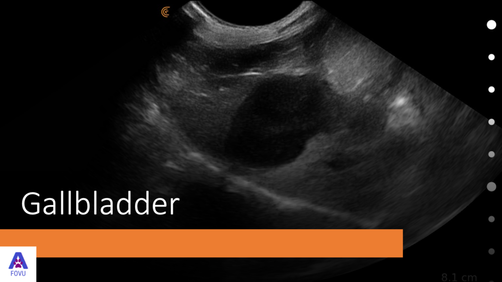
A gallbladder is a fluid-filled organ, which will appear anechoic/black on the ultrasound screen. In dogs, the gallbladder sits between the quadrant and the right medial liver lobes. And in cats, it sits between two parts of the right medial liver lobe. The gall bladder’s job is to store and concentrate bile received from the hepatic biliary ducts.

The size of the gall bladder varies depending on the length of time since the last meal. Before meals, the gallbladder will be fairly large. In these cases, the gallbladder wall is really thin. We expect it to measure under 0.1 centimeters in cats and under 0.2 centimeters in dogs.
Indications for Scanning the Gallbladder
We’ll decide to scan the gallbladder if we see increased liver values such as increased total bilirubin (ALKP/Tbil).
If the animal presents with some cranial abdominal pain, we often think of pancreatitis, but we should also include gallbladder disease on our differential diagnosis list. We can also use a gallbladder scan to aid in the assessment of anaphylaxis, if we see the halo sign.
Common Artifacts
- Slice thickness artifact – the ultrasound machine averages out what it’s seeing between the wall and the content of the gallbladder and makes it almost appear like there’s sludge within the gallbladder.
- Acoustic enhancement – as sound waves pass through fluid, they’re not attenuated so beyond the gallbladder, imaging will appear brighter.
- Refraction – we can see ultrasound waves bounce off the corners of the rounded surface of the gallbladder.
- Mirror image artifacts – when there’s gas in the lungs, we end up with a mirror image on the opposite side. That tells us the lungs are nicely full of gas.
Normal Ultrasound Views of the Gallbladder
Below is a sagittal view of the mirror image of both the liver and the gallbladder.

We fan through the liver and gallbladder. The gallbladder lies right of the midline. As we fan through the liver and gallbladder in this next video, the stomach just pops into view.

The gallbladder lies right of the midline. So, when we place the probe on sagittal leave, we won’t necessarily see it, we’ll need to aim the probe slightly to the right of the midline. Note that cats have this quirk – they can have bilobed gall bladders, which can be completely normal.
Common Pathology Seen in the Gallbladder
- The gallbladder can be large because there’s obstructive disease. You need to determine whether it’s intrahepatic or extrahepatic. Use ultrasound to investigate whether something is stuck in the lumen of the bile ducts.
- Look at the margins to see if there is wall edema or increased thickness of the wall, which we expect to be under 0.2 centimeters in healthy dogs.
- Check for free fluids due to a gallbladder rupture, which is extremely serious for the animal.
- Look at the contents of the gallbladder lumen to check for sludge – is it mobile or non mobile? Shadowing or non-shadowing?
- Watch for polyps, mucoceles, and choleoliths and note if it’s focal, multifocal, or diffuse throughout the gallbladder.
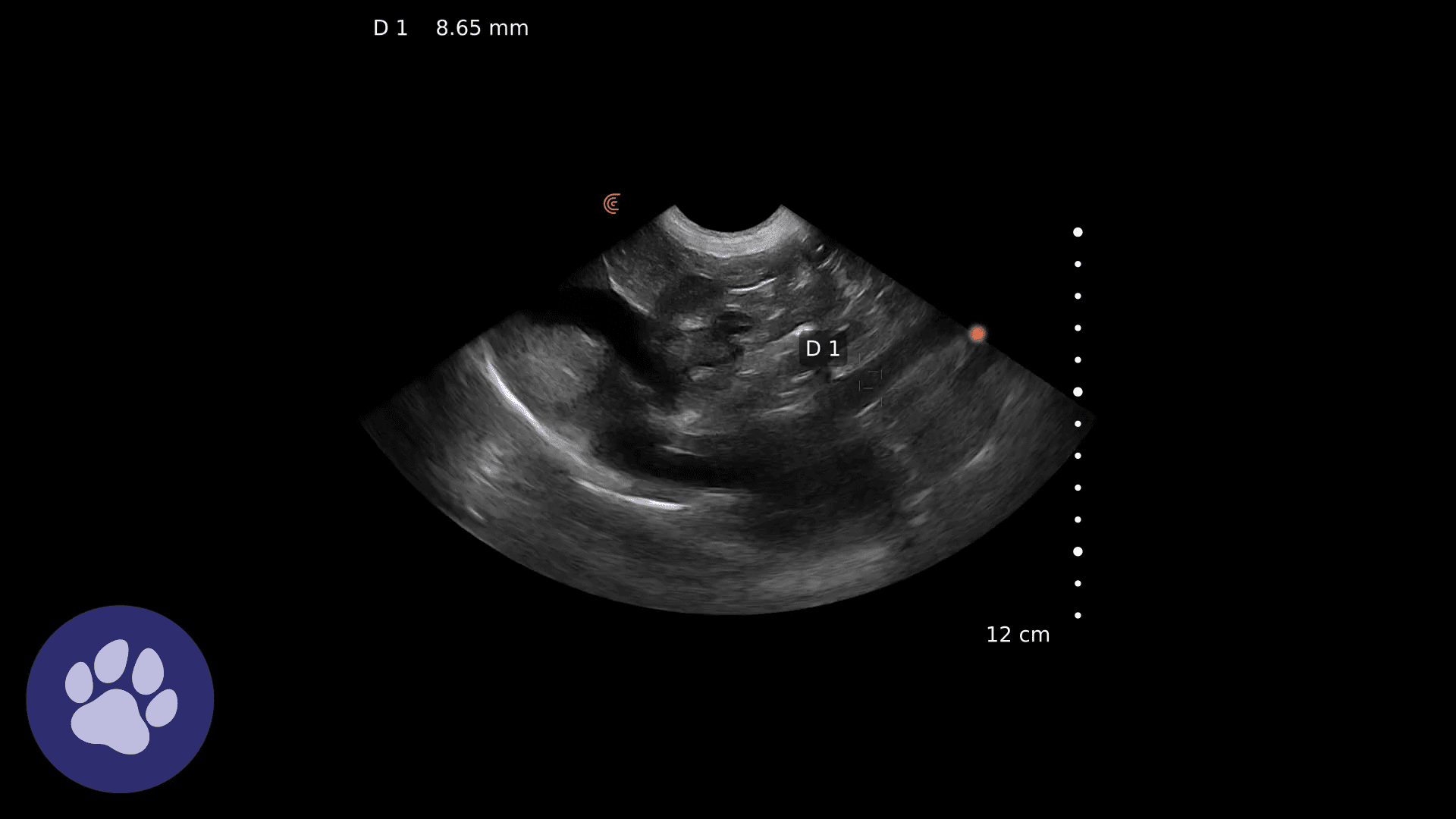
Video Clips: Sludge in the Gallbladder
In this next video, you see the sagittal view of a liver and a gallbladder with some sludge. The diaphragm is the bright white line.
Occasionally, you will can see the beating heart through the diaphragm. As the exam fans down towards the right, you see the gallbladder coming into view and you’ll see some sludge.
In this video, you see the gallbladder and again there’s some hyper echoic sludge that’s falling to one side. It’s falling to the gravity dependent side. If we were to flip the dog over, we would see it falling to the other side after a few minutes.
In this transverse view, you see the more horizontal diaphragm the gallbladder with a small amount of sludge.
Tips on Evaluating Gallbladder Sludge
Gallbladder sludge is not always significant and ultrasound is an essential tool when assessing sludge.
It can be normal in elderly dogs to find sludge, for example. So, we need to assess to see if it’s abnormal or not. In cats, it’s always abnormal, so if we find sludge, we know something’s going on in the gallbladder.
We can use ultrasound to see tortuous bile ducts. When bile is struggling to get through the bile ducts, they can become tortuous. Tortuous bile ducts can be normal in cats, but it’s definitely abnormal in dogs.
During an ultrasound exam, we can also see mineralization. On ultrasound, the sludge might be quite bright and look almost white. Then we’ll have an acoustic shadow so we can’t really see through it. And there may even be stones.
While we’re scanning, we need to assess the mobility of the sludge. Is it able to move from one side to another? And, do we have an intraluminal or extraluminal obstruction? Is there any gallbladder wall thickening? That can help us to confirm cholangitis.
Ultrasound of the gallbladder is important as it adds evidence to blood test results and the animal’s health history. It’s a really useful tool to use a series of scans to monitor treatment. For example, if an animal is ill with a cholangitis, it might need multiple scans over a period of months to assess when treatment can be stopped.
Repeat gallbladder exams over time are also useful to assess for disease progression. Sometimes we see an abnormal collection of mucus and bile salts, which can lead to biliary mucosal. These are really serious and can become a surgical emergency because they can lead to obstruction and necrosis and gallbladder rupture.
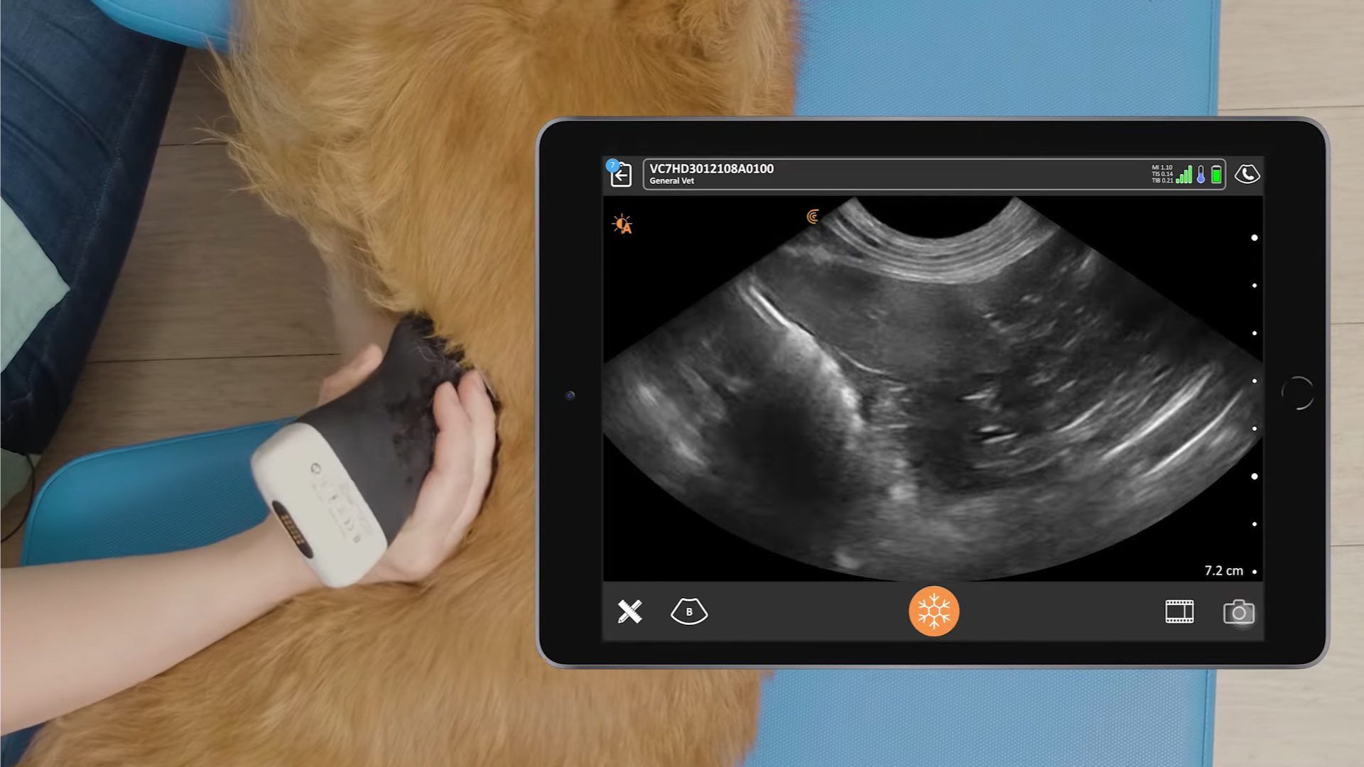
[VIDEO] How to Scan the Gallbladder on a Dog Using Ultrasound with the Clarius C7 HD Vet
Watch this 2-minute Clarius Classroom video to learn Dr. Edwards’ step-by-step procedure to scan the gall bladder of a dog to check for stones, sediment, sludge, and gas.
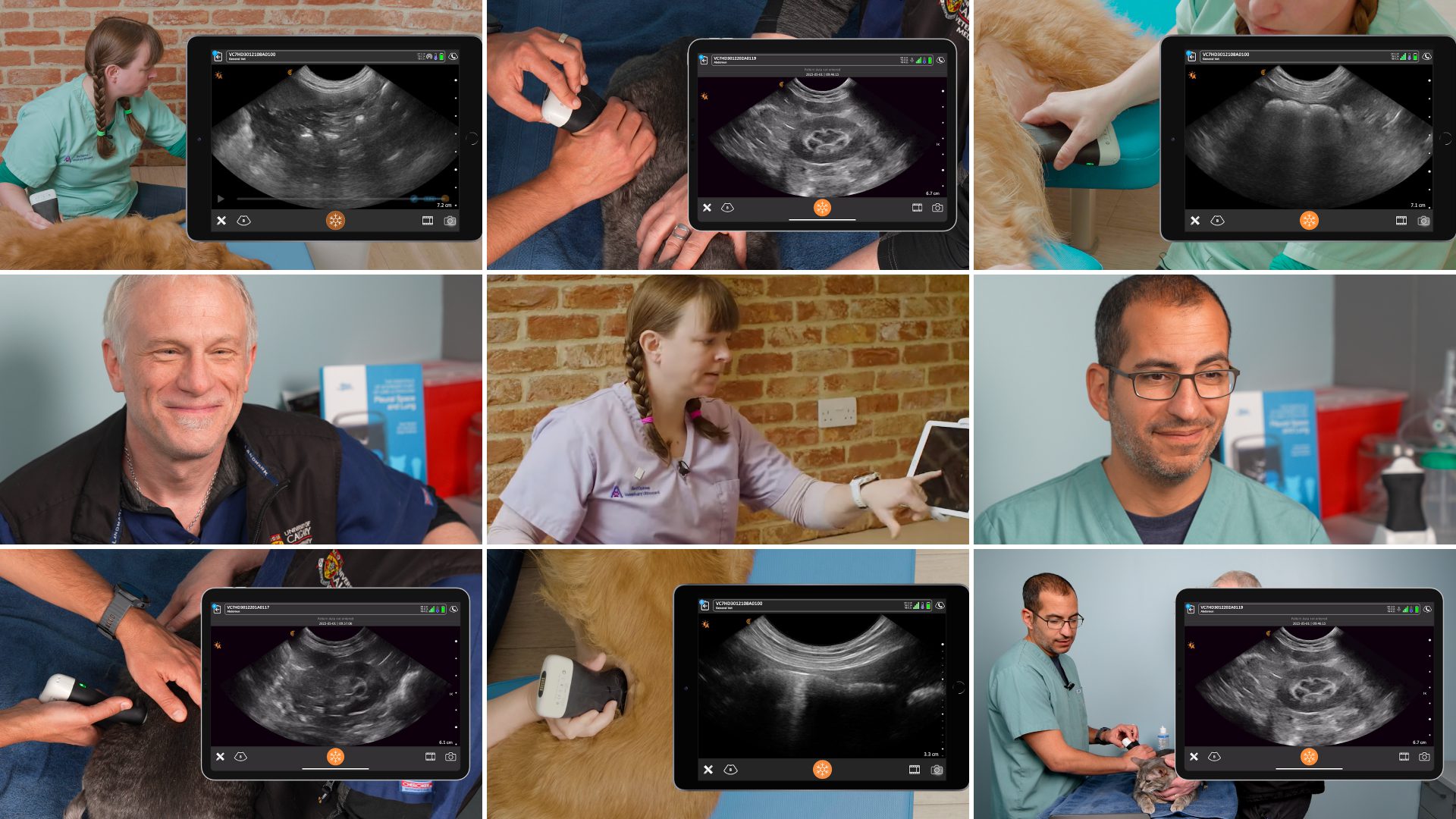
Want to Learn More? Watch this On-Demand webinar on PLUS Exams!
If you’re interested in learning more about veterinary ultrasound, watch for our fun, fast-paced webinar “Veterinary Point-of-Care Pleural Space and Lunch Ultrasound (PLUS) for Everyday Practice” featuring educators Dr. Soren Boysen, DVM, DACVECC and Dr. Serge Chalhoub, BSc, DVM, DACVIM (SAIM).
Clear Ultrasound Imaging You Can Trust for Your Veterinary Practice
With high-quality imaging and an AI-driven app that is almost as easy to use as your smart phone, Clarius is the ideal ultrasound system for veterinarians who are new to ultrasound.
To learn about how easy and affordable it is to add Clarius handheld ultrasound to your veterinary practice, visit our Clarius veterinary ultrasound page for a video demonstration and product details. Or contact us today to discuss which scanner is right for your veterinarian practice.
If you’re interested in alerts on new Clarius Classroom videos are they are published on Clarius Classroom, please subscribe to our Facebook, Twitter, LinkedIn, Instagram or YouTube channel.
You can also watch Dr. Edwards’ review of the Clarius C7 HD Vet on her website.


