Earlier this month, we introduced Clarius Classroom, a free online resource featuring instructional POCUS (point-of-care ultrasound) videos. One of our primary contributors is Dr. Oron Frenkel, an emergency physician, ultrasound educator, and Chairman of the Clarius Medical Advisory Board. We also invite specialist physicians to share their expertise.
This week, we’re sharing three short videos of lung pathology narrated by Dr. Frenkel and a 4-minute tutorial with Dr. Daniel Kim on how to scan the lungs.
Recognizing Pneumothorax on Ultrasound
Watch the following video to learn how to identify common signs of pneumothorax during an ultrasound exam.
Several pneumothorax signs are shown in this video clip captured with the Clarius L7 linear scanner.
- Pleural sliding is absent, as identified between the rib shadows.
- Corresponding M-mode shows the “bar code sign” where there is no change from superficial to deep to the pleural line, suggesting free air and lack of lung sliding.
- NOTE: while suggestive of pneumothorax, this finding can also be seen in bleb diseases or from pleural scarring.
Bright B-Lines May Signify Infection, Congestive Heart Failure, Pulmonary Fibrosis, or Scarring
In the following video, Dr. Frenkel shows us what B-Lines look like on ultrasound scans of the lung.
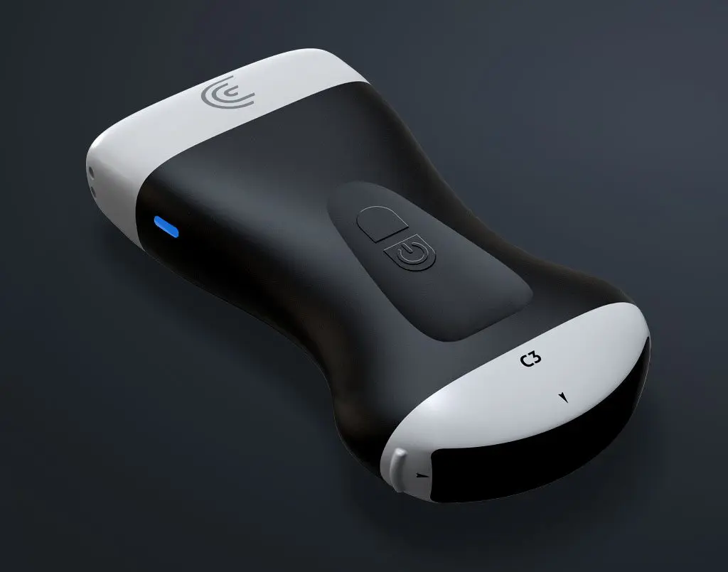
Here, B-Lines are captured with the Clarius C3 HD curvilinear ultrasound scanner, obscuring the A-Line pattern.
- Bright narrow beams that extend from pleural line down to the bottom of the screen are an artifact created by interstitial fluid.
- This finding, while sensitive, is not specific and can be present due to one or more of: infection, congestive heart failure, pulmonary fibrosis, or scarring.
What Pleural Effusion Looks Like on Ultrasound
Dr. Frenkel describes signs of pleural effusion on an ultrasound scan in this next video.
Complex pleural effusion is captured with the Clarius C3 curvilinear ultrasound scanner.
- The pleural effusion seen here is fibrotic, suggesting it’s subacute or chronic in nature.
- Pleural effusions can range from pure anechoic, to fibrotic, to frank iso- or hyperechoic fluid that is pathognomic for an exudate.
Lung Scanning Tutorial with Dr. Daniel Kim
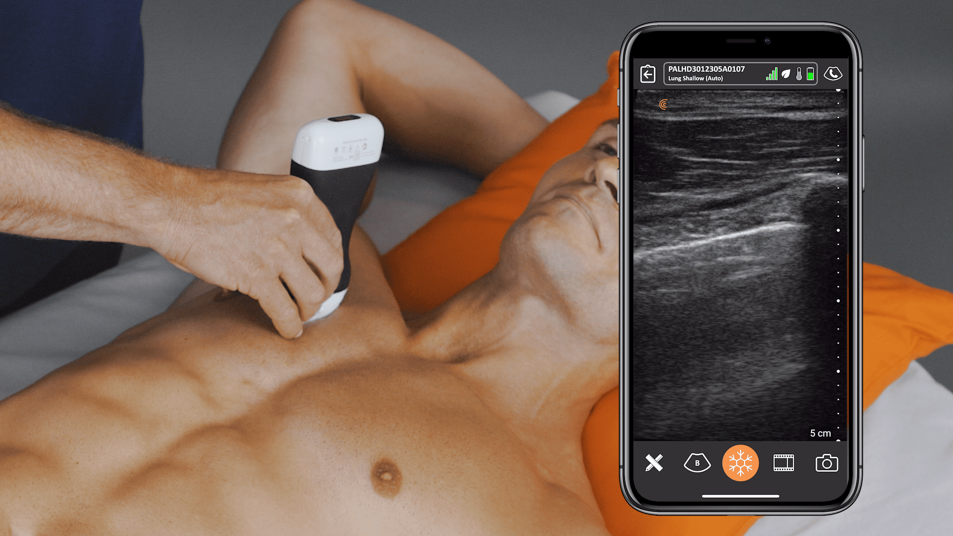
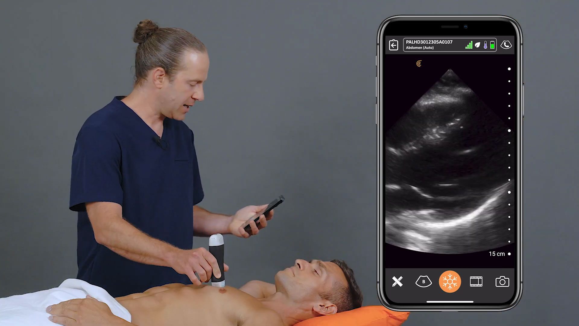
In any patient with dyspnea, tachypnea, or hypoxemia, lung ultrasound can rapidly and effectively diagnose the presence of interstitial fluid, larger consolidation, and pleural fluid. The degree of findings often correlates with severity. Watch this four-minute video to learn how to methodically scan the lungs with ultrasound.
Dr. Kim shows us how to interrogate the entire chest area using a six-zone scanning technique.
With high-quality imaging and an AI-driven app that is almost as easy to use as your smart phone, Clarius is the ideal ultrasound system for clinicians who are new to ultrasound. To learn more about how easy it is to learn and use the Clarius C3 multipurpose ultrasound scanner for point-of-care ultrasound exams, contact us today or request an ultrasound demo.
If you’re interested in seeing more ultrasound educational videos on Clarius Classroom as they are released, we invite you to subscribe to our Facebook, Twitter, LinkedIn, Instagram or YouTube channel for alerts.
