












Clarius Classroom

Anterior Knee Ultrasound with T-Mode™ and MSK AI

Dr. Christopher 'CJ' Miller

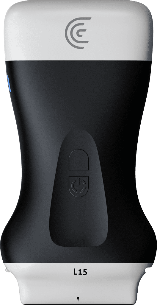
In this Clarius Classroom tutorial, physical therapist C.J. Miller from Frontline Function demonstrates how to utilize the advanced AI features—specifically T-Mode™ and MSK AI—of the Clarius L15 scanner to evaluate the anterior knee.

Key Highlights

Superior Knee & T-Mode™ Identification: The exam begins at the superior knee, utilizing T-Mode™ to automatically identify and label anatomical structures in real time, including the kneecap, quadriceps tendon, fat pads, and femur bone.

Color Opacity for Education: Miller highlights the ability to toggle color opacity over the identified structures, creating an intuitive visual aid that is highly effective for patient education and training new clinicians.

Trochlear View in Flexion: By moving the patient’s knee into full flexion, the practitioner evaluates the trochlear view to clearly observe the femoral cartilage, an ideal method for assessing conditions like osteoarthritis, gout, pseudogout, or general bony changes.

Distal Patellar Tendon & Hoffa’s Fat Pad: Moving distally, the scanner easily identifies the patellar tendon, patellar bone, and Hoffa’s Fat Pad.

Automated MSK AI Measurements: Switching to the MSK AI mode, the software automatically highlights the patellar tendon. Upon freezing the image, the AI instantly calculates and displays the thickest point of the tendon (measuring 4.8mm), bypassing the need for manual caliper placement and saving valuable time.
Specialties: MSK, Physiotherapy
Applications: Knee, MSK

00:00
–
00:11
My name is C.J. Miller, and I'm a physical therapist with Frontline Function. Today I'm going to be demonstrating the L 15 using its AI features of T mode and MSC AI on the anterior knee.
00:12
–
00:17
We'll start our exam at the superior knee. Looking at the quad tendon,
00:18
–
01:01
as well as the fat pads above the quad muscles and the kneecap. So I'm going to palpate the superior aspect of our patella. And I'll place my probe in long axis. And that should already give me a pretty good view of our quad tendon. Now the great thing about the T mode is that now I can turn it on and it will identify my structures for me.
So kneecap being on the top right of the screen, quad tendon coming down and inserting onto the kneecap. We've got our fat pads. And then the deepest part being our femur bone, I can scroll up and I can scroll down.
01:03
–
01:31
I can also turn on the opacity on the left side. And that will give me shades of color on each of these things as well, which is a really helpful piece. When you're educating other clinicians, but also if you were looking at this with a patient and they've never seen an ultrasound image before, you would be able to turn this on and they would immediately understand what they're looking at.
I'll turn the colors back off. I can also go into long axis mode,
01:32
–
01:45
and now I can identify the same things femur at the bottom of the screen, fat quad tendon as well as on the right. I'm going towards his vastus medialis muscle. As I follow this up,
01:47
–
01:55
it will track that tendon as well as be able to show me where the beginning of that vastus lateralis muscle comes in.
01:57
–
02:54
And then again, I can apply color on the left side to that. As I come down closer to the kneecap, I can move in him into full flexion. And now we can evaluate the. We call this the trochlear view of the anterior knee. This really highlights well the femoral cartilage. So you can see the cartilage labeled on the right side of the screen.
It also still shows our quad tendon overlying the cartilage. Our vastus medialis muscle and the bony groove of the femur. I can highlight that in a different way on the left side of the screen as well. This is a great area to evaluate for things like osteoarthritis, or for gout, or pseudo gout, or for cartilage changes, or just bony changes along the femoral condyles.
As I bring him back down into our starting position, I can come down to evaluate the distal patellar tendon.
02:56
–
04:35
I'll palpate first, find our tendon and place my pro in line with it in a long axis view. And now I can use T mode to automatically identify what's on my screen for me. So the left side of our screen is our patellar bone. We've got our patellar tendon coming across the screen and long axis and coming down and inserting onto the tibia.
Below that we have our Hoffa's Fat Pad, which is identified on the right side of the screen as fat. I can again turn on our colors here to identify these structures really well. So as we continue to look at the anterior knee with the patellar tendon and the patellar bone and Hoffa's fat pad. I can remove my T mode, and I can go to our MSK AI mode, and this is automatically going to highlight just the patellar tendon.
My favorite part about this is if I freeze, it will automatically give me a measurement at the thickest part of the tendon. So the AI will track the tendon, identify the thickest point, and at this on the top left you can see that this is about 4.8mm. This is saving me a lot of time. Usually I would have to freeze my screen, pull up my measurement tool, place my calipers on the exact area that it needs to go, and that's just a time saver to be able to click.
One button highlights the tendon for you and then gives you a measurement at its thickest part. So that's how we can use T mode in AI to evaluate the anterior knee. This is a really helpful tool when your patients are learning about ultrasound for the first time, and you're trying to educate them, it's also a really helpful tool.
If you were ever teaching musculoskeletal ultrasound to a clinician who's also trying to learn.

Products Used

More Classes

Request a Quote

With 8 scanners available, we offer a wireless ultrasound solution tailored to your needs. Request a quote to discover which scanner can deliver the best ultrasound imaging for your practice.

⚠️ Clarius ultrasound is for medical professionals only.

By providing my email, I consent to receive Clarius webinar invitations, case studies, whitepapers, and more, and I consent to the Clarius Privacy Policy. I can unsubscribe anytime.