While many aesthetic injectors still rely on tactile feel and generalized textbook diagrams, Dr. Roya Zarmehr Zamin, a global expert in ultrasound-guided aesthetics, represents a shift toward a more rigorous standard: visualized safety.
Dr. Roya advocates for the integration of point-of-care ultrasound (POCUS) as a practical, daily tool to navigate the highly variable and unpredictable vascular maps found in every patient. Rather than guessing the depth of a vessel or the location of previous filler, she uses high-frequency imaging to recognize the unique anatomy of each patient before the needle ever touches their skin.
Dr. Roya has developed repeatable FACT protocols enabling clinicians to move from blind techniques to a documented, evidence-based approach that optimizes practitioner confidence, patient safety, and outcomes. She shares her practical tips during a 1-hour webinar that is now available to watch on demand: Ultrasound for Facial Aesthetics: Practical Tips to Simplify Facial Ultrasound. Whether you’re beginning your ultrasound journey or looking to refine your skills, this webinar delivers detailed, actionable techniques you can apply immediately in your practice. Watch the webinar at your convenience and scroll down for highlights.
Pro Tips for Reproducible Results
Using ultrasound won’t make you a radiologist… it’s just to recognize the unique anatomy and injecting more safely,” says Dr. Roya.
Her FACT approach for aesthetic clinicians focuses on making ultrasound reproducible and simple enough for a high-volume clinical environment. To make ultrasound a seamless part of your daily workflow, Dr. Roya recommends several practical maneuvers:
- Vascular Mapping is Mandatory: Never assume an artery is exactly where a textbook suggests; map high-risk zones to identify vessel depth and location before injecting.
- The “Zero Pressure” Technique: Use a thick layer of gel to fill facial “hills and valleys,” gliding the scanner over the gel rather than the skin to avoid compressing vessels or distorting measurements.
- The “Pen Grip” vs. “Palmar Grip”: Use a pen-style grip for superficial structures and a palmar grip for deeper areas like the temple or jawline.
- Anchor Your Hand: Always use your fourth or fifth finger as an “anchor” on the patient’s face to stabilize the image.
- Dynamic Assessments: Identify muscles like the masseter, frontalis, or buccinator by asking the patient to move or clench, allowing you to visualize contractions and tissue planes in real-time.
VIDEO Demonstration: Safe Mapping of Vascular Anatomy
Watch this 12-minute video to see Dr. Roya demonstrate facial mapping in key areas prior to treatments using the Clarius L20 HD3 ultra high frequency wireless ultrasound scanner.
Identifying “The Shades of Gray”: Filler Signatures
Recognizing how different fillers appear on ultrasound is vital for both placement and complication management.
- Hyaluronic Acid (HA): Typically appears as black (anechoic) pockets, often with “posterior enhancement” or brightness underneath.
- Calcium Hydroxyapatite: Shows as bright white (hyperechoic) with “posterior shadowing” because the material blocks the ultrasound beam.
- Permanent Fillers (e.g., Silicone): Often produce a “snowstorm” pattern or “comet tail” artifacts that obscure underlying tissue layers.


Watch this webinar to see Dr. Roya demonstrate in detail how to differentiate these fillers in real-time.
Why Experts Choose Clarius for Aesthetics
During the webinar, Dr. Roya and the Clarius team highlighted several features that make the Clarius L20 HD3 the premier choice for aesthetic clinicians:
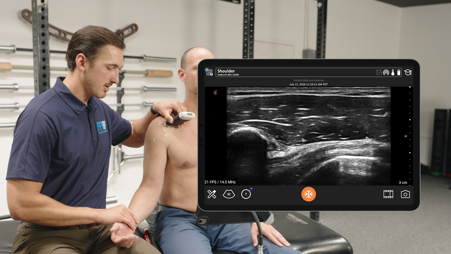
- Ultra-High Frequency: With up to 20 MHz, it provides the specialized resolution required to see the finest details of the skin and superficial vessels.
- Wireless Freedom: The scanner is 30% smaller and lighter, with no wires to sterilize or trip over.
- Automated Presets: Specialized settings for the temple, chin, lips, and more automatically optimize the gain and depth for that specific area.
- Advanced Guidance: Specialized needle enhancement and pulse Doppler tools help you ensure you are “not injecting what you cannot see”
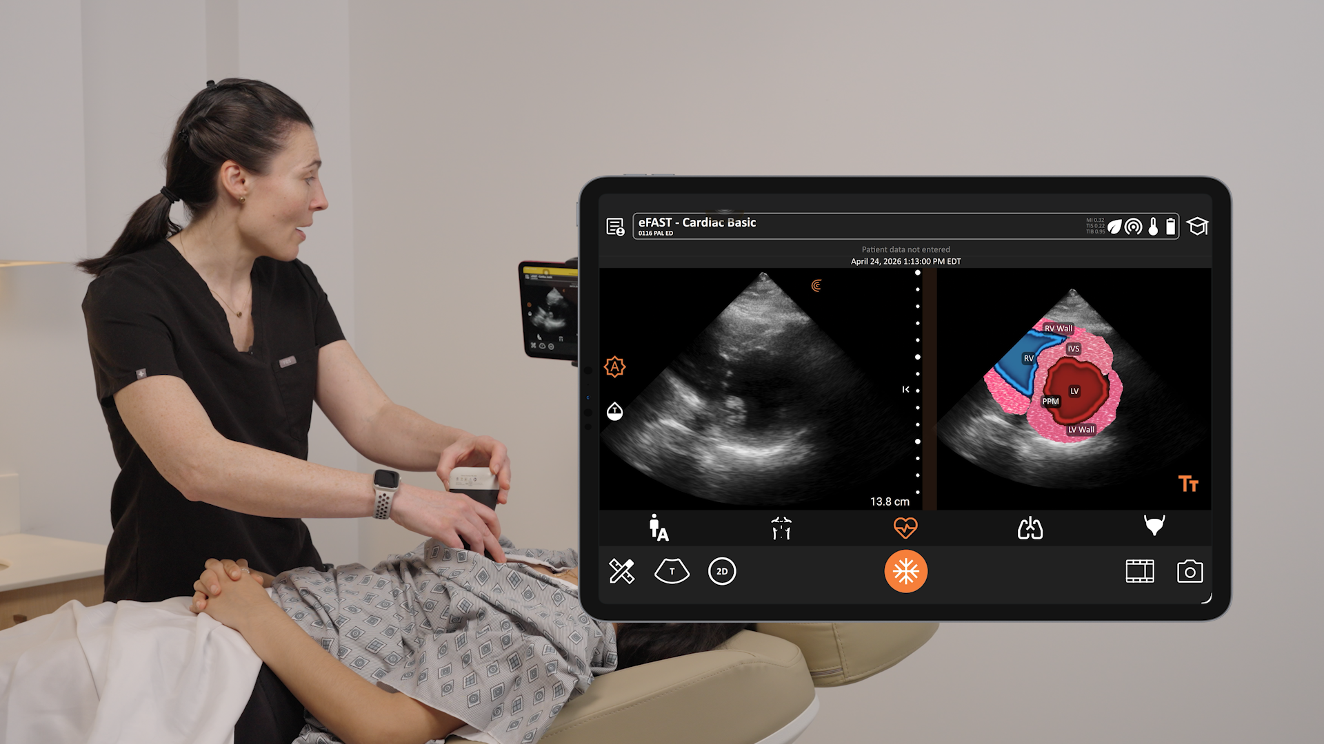
- T-Mode™ Technology: AI-powered T-Mode™ overlays distinctive colors, patterns, and labels to instantly identify and differentiate tissue layers in the face, which is helpful for aesthetic practitioners learning how facial anatomy works and where to target injections. Watch this 2-minute video to see it in action.
Learn more about Clarius AI-powered ultrasound on the aesthetics specialty page. Or contact us for a personalized virtual demonstration.