1
00:00:00,030 --> 00:00:04,510
Hello community, how are we today. Enrique Villanueva, musculoskeletal interventional radiologist
2
00:00:04,510 --> 00:00:10,870
and soft tissue specialist as an advisor for Isaacs. In recent years and since
3
00:00:10,870 --> 00:00:17,390
2018 we have learned the aesthetic view of plastic surgeons and what they really need
4
00:00:17,390 --> 00:00:23,750
to be able to perform a personalized anatomical marking. We have evolved that to
5
00:00:23,750 --> 00:00:30,110
be able to understand then what diastasis of the Linea Alba is. And something very innovative which is the diastasis
6
00:00:30,190 --> 00:00:35,590
of the semilunar lines, something that was not described and that we can see by ultrasound now,
7
00:00:35,590 --> 00:00:42,230
How to really mark pre and post surgery? How to evaluate the cellular tissue?
8
00:00:42,710 --> 00:00:48,990
Where is Camper? Where is Scarpa in liposuction? All this we can understand
9
00:00:48,990 --> 00:00:55,989
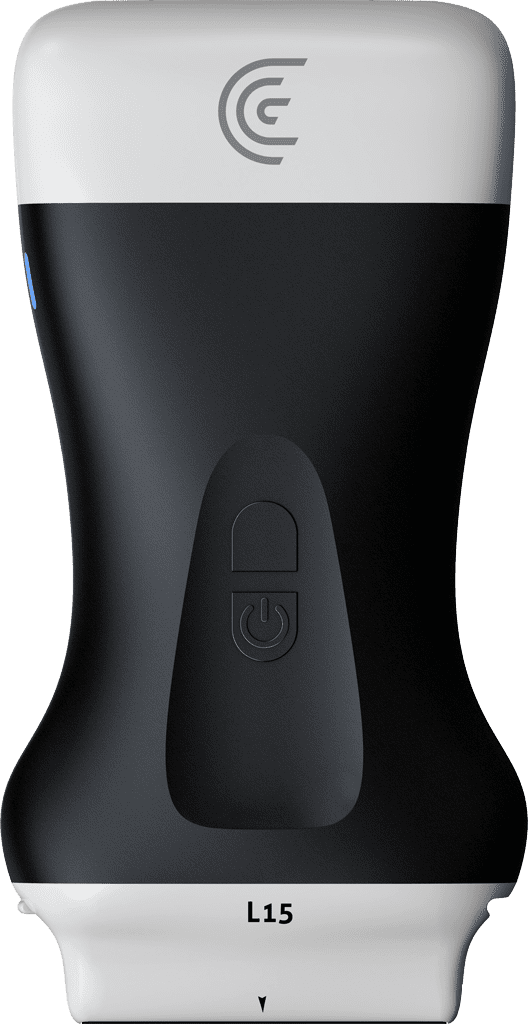
using a high resolution ultrasound such as the L15 or a Clarius L20.
10
00:00:56,150 --> 00:01:02,320
In this way we can also. If you do grafting, evaluate the vascular pedicles to
11
00:01:02,320 --> 00:01:09,000
be able to do the fat graft. In this episode we are only going to see how to evaluate the
12
00:01:09,000 --> 00:01:15,920
linea alba, the rectus abdominis and how you can mark all the part of the abdominal
13
00:01:15,920 --> 00:01:22,800
wall to be able to do your anatomical and high precision marking procedure with the
14
00:01:22,800 --> 00:01:28,839
ultrasound. Important then in abdominal wall to be able to identify that in plastic surgery and in
15
00:01:28,839 --> 00:01:34,959
abdominal wall procedures we divide the abdominal wall into supraumbilical and infraumbilical.
16
00:01:34,960 --> 00:01:40,720
We are going to work and mark a bit everything that is supraumbilical, since there are very important points
17
00:01:40,720 --> 00:01:47,720
like the Linea Alba. We are going to see the rectus abdominis, semilunar, external oblique,
18
00:01:47,760 --> 00:01:54,520
diastasis of the semilunar line. And we are also going to see if there is NOA fat which is the
19
00:01:54,520 --> 00:02:00,760
NO FAT, which is that retroperitoneal fat. And we are going to identify then the anterior and
20
00:02:00,760 --> 00:02:06,969
posterior belly of the rectus abdominis where you can inject. We are going to identify if there is Camper
21
00:02:07,009 --> 00:02:13,569
Scarpa in this procedure and the thickness of the subcutaneous cellular tissue. So in the midline
22
00:02:13,609 --> 00:02:20,570
we are going to start with our L15 transducer. As always, please. One centimeter
23
00:02:20,570 --> 00:02:27,130
below the line or surface anatomy reference in abdominal wall is the peritoneum.
24
00:02:27,449 --> 00:02:33,729
It is important that we always locate the peritoneum and there we are going to leave one centimeter of
25
00:02:33,729 --> 00:02:40,529
depth. In this way we are going to be able to have a certainty of what we are evaluating and
26
00:02:40,529 --> 00:02:45,210
we are going to be able to then identify all the structures that we want to evaluate. We begin supraumbilical
27
00:02:45,210 --> 00:02:51,689
we see how our left and right rectus abdominis looks, we see how
28
00:02:51,809 --> 00:02:58,689
the distance of our Linea Alba looks at this level. And
29
00:02:58,729 --> 00:03:05,219
it is important that for surgery if you want to do a very high definition surgery,
30
00:03:05,300 --> 00:03:11,819
a complete body definition the linea alba or the average over 2.5
31
00:03:11,820 --> 00:03:18,699
centimeters, the distance over 2.5 centimeters. That is called diastasis. Ready? Here
32
00:03:18,699 --> 00:03:24,779
we are going to measure the averages then. We continue in the middle third. We see our linea alba again.
33
00:03:24,820 --> 00:03:31,699
We measure again. Here we have 15.4 millimeters. We continue going up and here
34
00:03:31,699 --> 00:03:38,059
we are already going to the subxiphoid area. We see on the right side and on the left side how the
35
00:03:38,060 --> 00:03:44,659
costal insertion of the rectus abdominis is already seen and we see a very
36
00:03:44,700 --> 00:03:51,419
small linea alba of less than 3.3 millimeters. In this way we already reviewed our
37
00:03:51,420 --> 00:03:57,900
Linea Alba. Here we see at the subxiphoid level how there is a thickening of the retroperitoneal
38
00:03:57,900 --> 00:04:04,049
fat or the fat called. No fat. That is important because if you are going to do
39
00:04:04,049 --> 00:04:09,809
a liposuction, lipo-marking and are going to define the contour of the patient, it is likely that she will also
40
00:04:09,809 --> 00:04:14,969
go to nutrition and change habits. And this fat is a fat that has an important metabolic context
41
00:04:14,970 --> 00:04:21,649
Now we go and return supraumbilical and we are going to analyze the
42
00:04:21,649 --> 00:04:28,609
subcutaneous cellular tissue. And never stop looking at your Linea Alba, because the Linea Alba can
43
00:04:28,609 --> 00:04:34,289
have a solution of continuity and that is a gastric hernia, evaluate it always dynamically.
44
00:04:34,289 --> 00:04:40,329
We can tell the patient to push out the abdomen or to inflate the abdomen or to
45
00:04:40,329 --> 00:04:46,329
use force to contract and you will see how it bulges through there our hernias. In this case we do not
46
00:04:46,329 --> 00:04:53,329
have it subcutaneous tissue. We are going to measure the subdermal space to the fascia of the
47
00:04:53,329 --> 00:04:58,849
Linea Alba and this is our thickness of the subcutaneous cellular tissue. You can evaluate the
48
00:04:58,849 --> 00:05:04,579
thickness of the subcutaneous cellular tissue of the entire abdominal wall and in that way you can
49
00:05:04,579 --> 00:05:10,659
do a qualitative and quantitative control of the thickness of the subcutaneous cellular tissue and be able to
50
00:05:10,659 --> 00:05:17,059
evaluate during the surgery and after the surgery, what is the final thickness that this
51
00:05:17,059 --> 00:05:23,699
fat has. It is important and we are averaging that an adequate healthy thickness is
52
00:05:23,700 --> 00:05:30,379
at least 1.5 millimeters of thickness below the skin. That would be the
53
00:05:30,380 --> 00:05:37,219
ideal so that we can have a fat that metabolically generates security
54
00:05:37,219 --> 00:05:43,578
for the patient. We continue observing then the subcutaneous cellular tissue and as you can see, two
55
00:05:43,579 --> 00:05:50,339
hyperechoic lines are seen at the level of the subcutaneous cellular tissue. These two hyperechoic lines
56
00:05:50,380 --> 00:05:56,980
one and two are Camper and Scarpa, and everything that is below the
57
00:05:56,980 --> 00:06:03,939
Scarpa is deep fat. Many surgeons around the world do Deep Plane. Liposuction.
58
00:06:03,980 --> 00:06:10,579
And it is important to know that thickness. In that way they will be able to understand what technology they can
59
00:06:10,579 --> 00:06:17,379
use if they do not use technology or how much liposuction to be able to do the lipo-marking in that area
60
00:06:17,379 --> 00:06:24,299
your patient requires. In this way we have evaluated midline important
61
00:06:24,299 --> 00:06:30,499
and do not forget in the midline and periumbilical. We have to identify what I call
62
00:06:30,499 --> 00:06:36,539
masses of the abdominal wall. The masses of the abdominal wall with the boom of the Ozempic. We have to
63
00:06:36,540 --> 00:06:42,819
identify if there are tracts and thickening of the subcutaneous cellular tissue associated with injections
64
00:06:42,820 --> 00:06:49,500
of fat tissue, fat tissue, like in the PICs or insulin dependents. And if they are
65
00:06:49,500 --> 00:06:56,379
women, we always have to infraumbilical identify if it is possible or probable that these
66
00:06:56,380 --> 00:07:02,499
lesions in subcutaneous cellular tissue have to do with endometriosis. So anatomical awareness
67
00:07:02,499 --> 00:07:08,879
also has to do with pathological anatomical awareness. Let's look for endometriosis,
68
00:07:08,920 --> 00:07:14,159
which probably that could be one of the causes of weight changes in your patient. And it is
69
00:07:14,160 --> 00:07:20,239
important because you sometimes in surgery think it is a fibrosis now in midline
70
00:07:20,239 --> 00:07:26,480
supraumbilical. I go periumbilical and I stay with a rectus abdominis. Look how rectus
71
00:07:26,480 --> 00:07:33,239
abdominis looks with transverse traces. If we are transverse, I turn clockwise
72
00:07:33,240 --> 00:07:39,920
and there we are going to observe our metamer. Look how the transition looks. There
73
00:07:39,920 --> 00:07:45,319
we see an echogenic line that divides the rectus abdominis into superior belly and inferior belly.
74
00:07:45,359 --> 00:07:52,199
I'm going to go cephalad, yes, and there we're going to see how there's a transition and we see
75
00:07:52,199 --> 00:07:58,000
our first transition. And that is a metamer. So, what do we do in the midline?
76
00:07:58,040 --> 00:08:04,910
We can mark our first metamer. In this way we are going to be able to start making
77
00:08:04,910 --> 00:08:11,869
anatomical marking. We continue cephalad, rectus abdominis in longitudinal.
78
00:08:11,870 --> 00:08:18,389
And look, here our metamer appears again in the midline. And there then we
79
00:08:18,430 --> 00:08:25,349
mark again where metamer number two is located. In this way we are going to
80
00:08:25,349 --> 00:08:31,828
start looking for the metamer. Remember, we begin periumbilical. I see our rectus
81
00:08:31,829 --> 00:08:37,710
abdominis, we rotate clockwise, we see how the superior belly and inferior belly of the rectus
82
00:08:37,710 --> 00:08:43,669
abdominis are. I go cephalad and there I begin to see the rectus transitions. And
83
00:08:43,669 --> 00:08:49,869
those are the metamers. Perfect, now I return. Periumbilical.
84
00:08:50,390 --> 00:08:56,669
I go towards the outer face, towards the flank and look how we are going to see this second transition and this
85
00:08:56,670 --> 00:09:03,349
transition between rectus abdominis, transversus, internal oblique and external oblique is what
86
00:09:03,450 --> 00:09:10,169
forms our semilunar line. An important tip. If you inject and do a
87
00:09:10,169 --> 00:09:16,809
block between the transversus and the internal oblique, it is a total block and that can improve the
88
00:09:16,809 --> 00:09:23,489
surgical experience of your patient. Now, do not forget that in your L15 ultrasound
89
00:09:23,650 --> 00:09:30,608
we also have an augmented reality mode, an artificial intelligence mode
90
00:09:30,609 --> 00:09:36,009
where it can help you understand the anatomical structures, mainly in
91
00:09:36,010 --> 00:09:42,530
patients who perhaps have sarcopenia and that we have a structure very mixed between the
92
00:09:42,530 --> 00:09:49,169
fat and between the muscles. This is one of the advances of how we can mark the abdominal wall
93
00:09:49,169 --> 00:09:55,609
in the midline. Remember to evaluate diastasis hernias. Superficial fat masses,
94
00:09:55,610 --> 00:09:57,010
deep fat.