For decades, the standard pathway for a patient with suspected Carpal Tunnel Syndrome (CTS) has been a lesson in patience: a clinical exam, a referral out for nerve conduction studies, a two-week wait for an appointment, and a follow-up visit to finally discuss surgery.
According to Dr. John R. Fowler, an orthopedic surgeon at the University of Pittsburgh, that model is officially outdated.
I personally no longer routinely order nerve studies for carpal tunnel workup,” says Dr. Fowler. “My evaluation has become much more efficient and reliable since starting to use the Clarius ultrasound system.”
For clinicians who feel intimidated by the “knobology” of traditional cart-based ultrasound, Dr. Fowler’s experience offers a roadmap for a more streamlined, patient-centric, and profitable practice. Watch this video to learn more about his experience with Clarius ultrasound. Or scroll below for highlights.
The “Garden Hose” Effect: Visualizing the Diagnosis
One of the biggest hurdles for patients is understanding why they need surgery. Dr. Fowler uses the Clarius scanner to provide immediate visual proof.
I talk about the old Looney Tunes cartoons, when they would step on a hose and the hose would kind of back up and blow up on the character. That’s what happens with carpal tunnel, and I can show them that nerve swelling in real-time.”
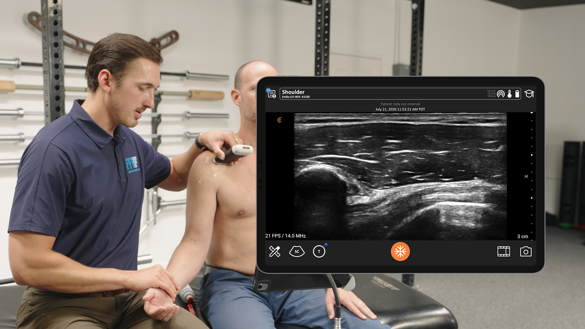
By scanning his own wrist with Clarius (typically 8 mm2) and comparing it to a patient’s swollen nerve (perhaps 15 mm2), Dr. Fowler creates a “seeing is believing” moment that builds instant diagnostic confidence.
The nice thing is, once you do a lot of these, it only takes about 15 or 20 seconds; doesn’t change your flow very much,” he explains.
Breaking the Learning Curve with AI
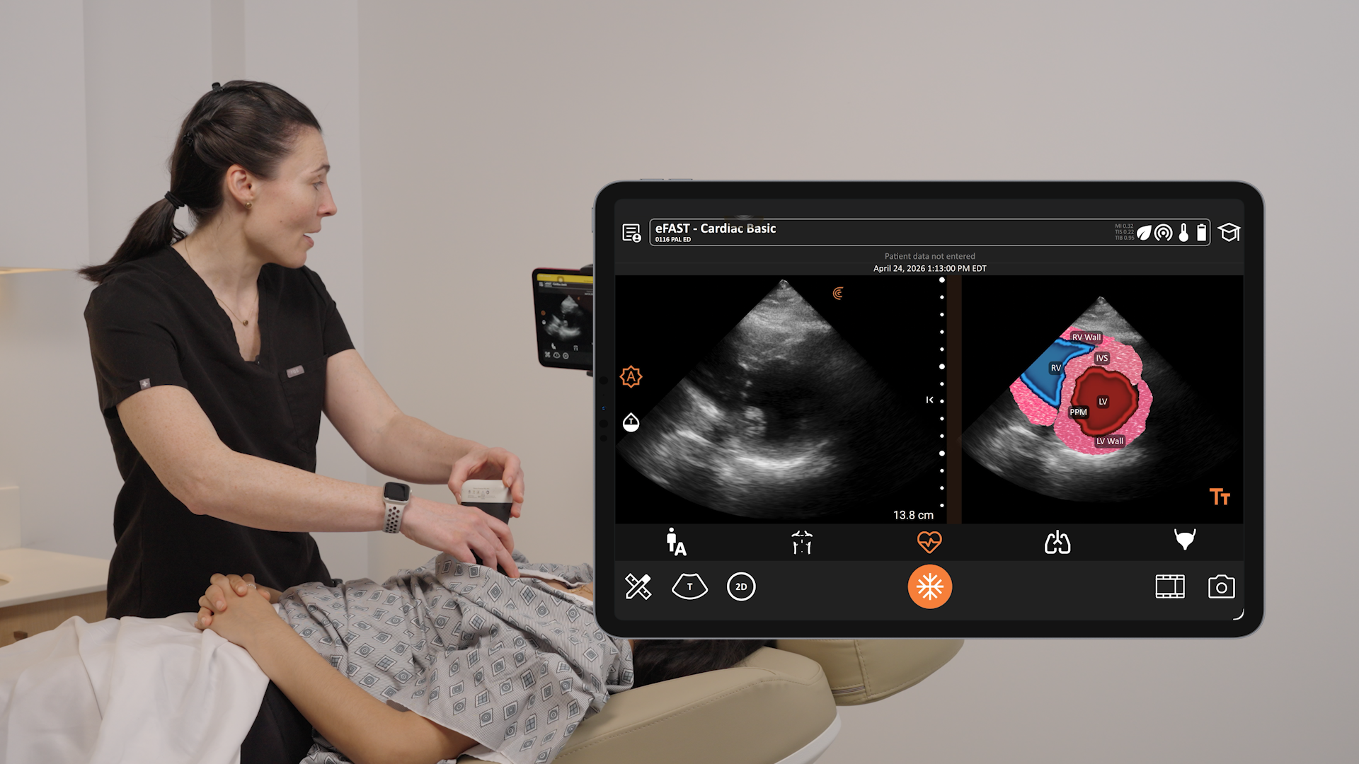
The most common reason MSK clinicians avoid ultrasound is the perceived difficulty of getting accurate measurements. Dr. Fowler has solved this by utilizing Clarius Median Nerve AI.
The AI automatically identifies the median nerve and calculates the cross-sectional area. It’s so reliable that Dr. Fowler now has his medical assistants perform the scan as a “fifth vital sign”—much like taking blood pressure—before he even enters the room.
- Ease of Use: “It was ridiculously easy… it maybe takes 2 or 3 minutes to learn how to do it,” says Fowler.
- Objectivity: AI removes human bias, scanning the nerve to find the maximum cross-sectional area more accurately than the human eye might.
- Delegation: Because the AI standardizes the measurement, even non-experts can produce reliable, surgical-grade data.
Video Demonstration: Median Nerve AI Watch this 2-minute video demonstration of Median Nerve AI, a tool used to locate and measure the median nerve in Carpal Tunnel Syndrome assessments.
The Bottom Line: Efficiency and ROI
Whether you are in a private clinic or a high-volume hospital, the operational impact of switching from “Cart to Handheld” is transformative.
1. Goodbye to the Cart (Operational Speed)
Traditional ultrasound systems are often bottlenecks. “Before Clarius, I’d have to drag a large cart-based machine into the room, plug it in… the batteries were never charged. That took 3 to 5 minutes just to get ready,” says Fowler. “Clarius is always ready.”
2. The Single-Visit Diagnosis (Patient Throughput)
In a traditional workflow, patients often leave the office only to return weeks later. Dr. Fowler’s research shows that using point-of-care ultrasound (POCUS) saves an average of 1.8 medical visits and 27 days of waiting. For the surgeon, this means faster clinical decisions and higher patient satisfaction.
3. Strategic Billing (For Practices in the United States)
For those in RVU-based systems, ultrasound acts as a force multiplier. Instead of just billing a standalone scan (0.3 Work RVUs), personally interpreting the scan increases the “Data Complexity” of the visit. This often allows a surgeon to shift a standard Level 3 office visit to a Level 4 gaining roughly an entire RVU per patient.
By streamlining the diagnostic pathway, surgeons in the United States aren’t just saving time—they are maximizing the value of every minute spent in the exam room.
Dr. Fowler’s Message for Hand Surgeons
According to Dr. Fowler, while there are roughly 4,000 hand surgeons in the U.S., many still rely on “false negative” prone nerve studies. He sees the tide turning.
We shouldn’t be referring out for ultrasound; we should be doing it ourselves. The learning curve is small, and the power of having that data in the hands of the surgeon is invaluable.”
Deepen Your Clinical Expertise: Ultrasound for CTS
Ready to master median nerve imaging? We invite you to watch the exclusive Clarius webinar, Ultrasound for Carpal Tunnel Syndrome.
Whether you are aiming to enhance your diagnostic confidence, explore the latest AI-driven measurement tools, or safely guide MSK interventions, this session is packed with practical insights you can bring straight to your clinic.
Gain More Insights Without the Wait
Clarius high-definition MSK ultrasound enables clinicians to diagnose injuries, guide procedures, and deliver confident results at the point of care. Request a virtual clinical demo today to discover why Clarius is the preferred choice for clinicians who demand excellence without compromise.