


Achilles Ultrasound Exam Review and Patient Education
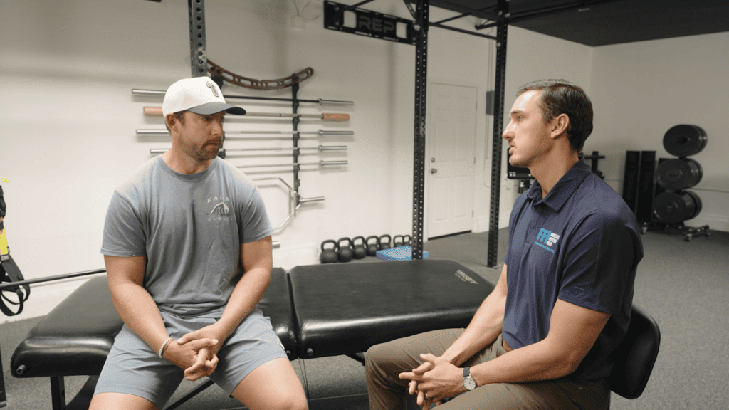
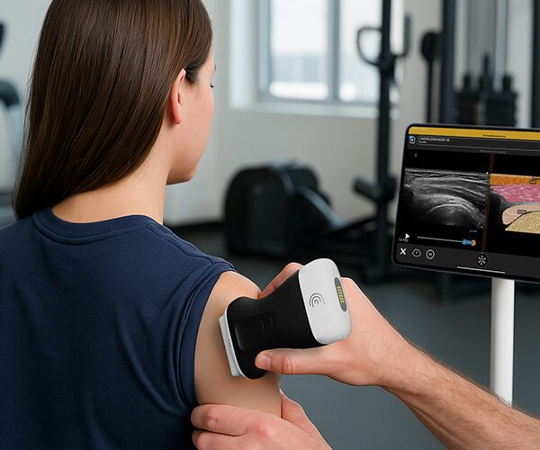
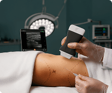
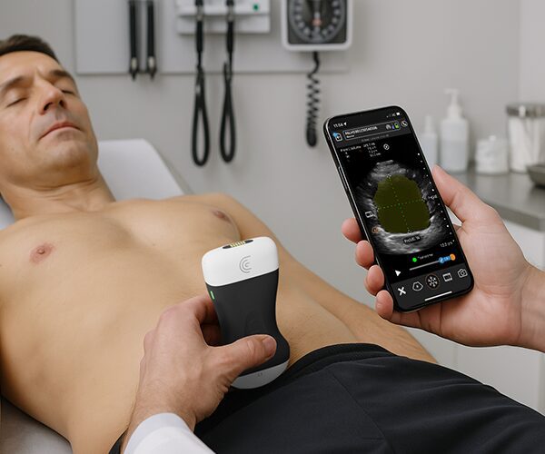
This video captures the conclusion of a physical therapy appointment where musculoskeletal ultrasound is integrated with a subjective history and clinical exam to educate the patient and develop a treatment plan.
Key Highlights
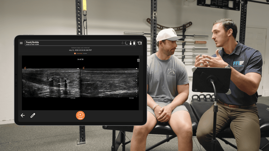
Immediate Patient Feedback: The practitioner reviews the ultrasound results live with the patient, Mike, providing on-site imaging feedback to save time and bypass the wait for external MRI or X-ray referrals.
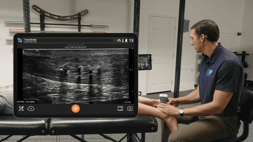
Side-by-Side Comparison: The ultrasound images contrast the patient’s left surgical Achilles tendon, which is thickened to over 12mm and contains visible anchors, against the right nonsurgical side, which measures a normal 5.5mm.
Diagnostic Reassurance: Despite the patient experiencing pain on the right side, the ultrasound confirms healthy tissue integrity, clean bone insertion, and an absence of significant Doppler blood flow. The practitioner explains this indicates early tendinitis rather than severe inflammation or tissue damage.
Rehabilitation Strategy: To promote healing, the practitioner prescribes eccentric loading exercises, such as backwards walking on a treadmill or slow-lowering reverse calf raises on a step, to load the tendon while it lengthens.
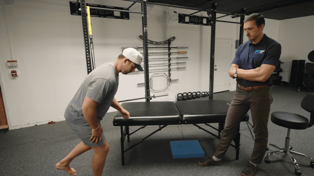
Return to Activity: Mike is instructed to let pain guide his running and hiking distances, incorporate single-leg balance exercises on various surfaces to improve right ankle stability, and safely return to controlled basketball activities, like shooting free throws, while avoiding full pickup games.