Dr. Joshua Guttman, POCUS educator and emergency medicine physician, gave the Clarius PAL HD3 an independent review. His verdict covers the AI features, the workflow, and what it all means for clinicians who want to scan with more confidence.
Dr. Joshua Guttman did not approach this review as a product demo. He carried the Clarius PAL HD3 through real emergency department shifts, scanned real patients with real pathology, and evaluated every feature against the demands of day-to-day clinical use.
As the founder of Peachtree POCUS, a nationally recognized POCUS educator, and a contributor to Rosen’s Emergency Medicine, Dr. Guttman has evaluated ultrasound devices with a specific lens: not what a device can do in a controlled setting, but what it does when a clinician is moving room to room, managing a full patient load, and needs answers fast.
His overall verdict on the Clarius PAL HD3: a strong, well-rounded device that stands out most for its AI integration and practical workflow design. Here is what he found, feature by feature.
This is a device that really leans into being both a clinical and an educational tool. That combination is genuinely useful.” — Dr. Joshua Guttman
Before the Scan: Real-World Durability and Workflow
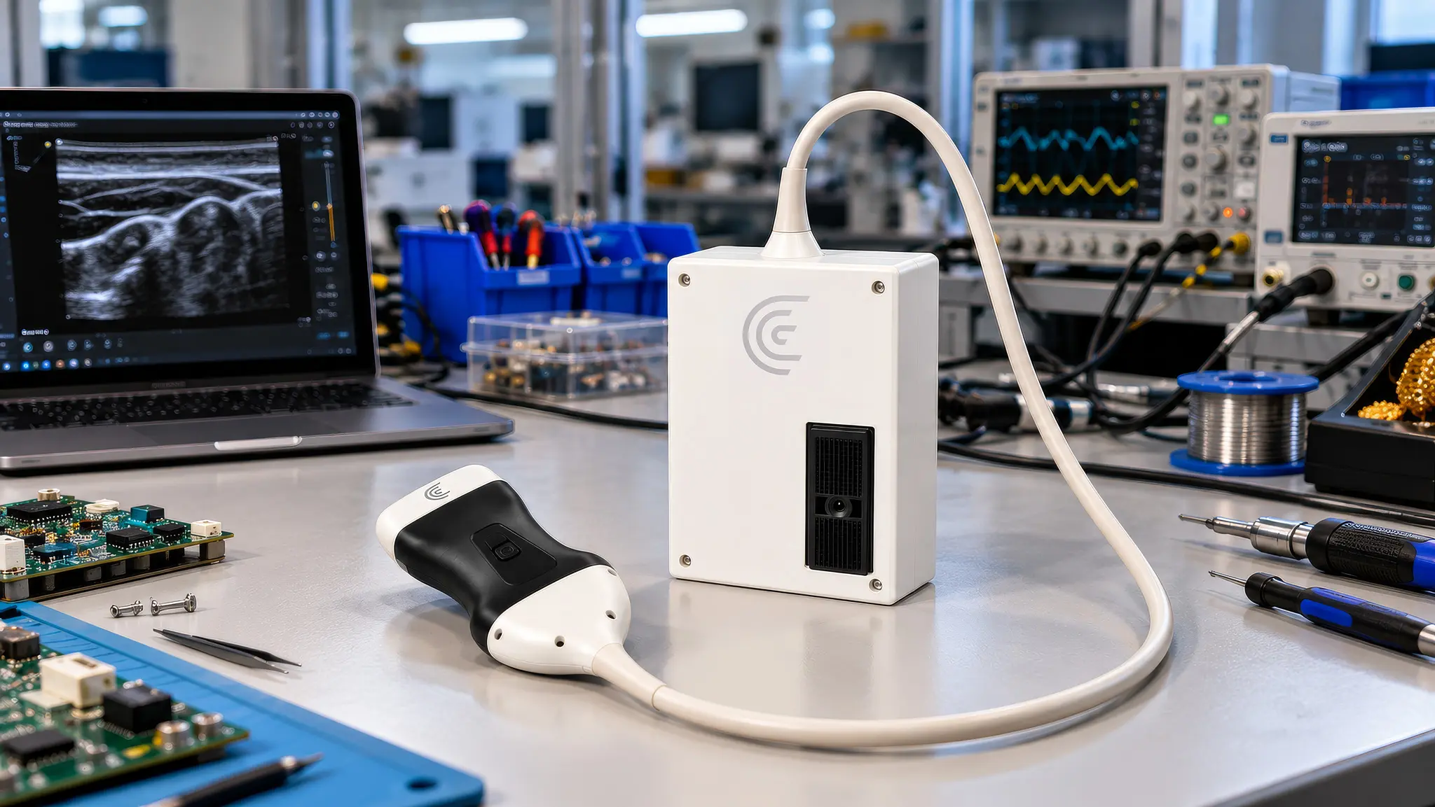
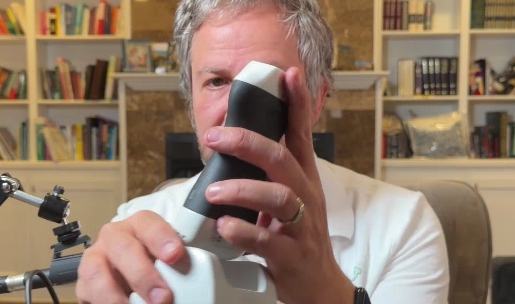
The Clarius PAL HD3 is a wireless, single universal scanner that handles curvilinear, phased array, and linear imaging without switching devices. That matters the moment you walk into a patient’s room carrying everything you need in one hand.
Dr. Guttman notes the probe is on the heavier side, with a wider footprint compared to smaller handheld devices. In real clinical use, though, the weight was never a limiting factor. He carried it through full ED shifts in what he describes as a “skinny pack,” moving from room to room without it slowing him down.
Heat and battery life are two of the most common real-world complaints with wireless ultrasound devices he notes. On both counts, Dr. Guttman found no meaningful issues. The battery held through a full shift of heavy scanning. Overheating was not a concern.
The Charging System That Gets out of Your Way
One detail Dr. Guttman specifically calls out: the dock-based charging system. No cables into the probe. No deliberate pauses to plug anything in. When he needed to top up mid-shift, he set the probe on the dock between patients and kept moving.
It’s one of those small things that actually makes a big difference in the day to day.” — Dr. Joshua Guttman
Clarius EF AI: Cardiac Confidence Without the Guesswork
The question physicians often face: Is this patient’s heart squeezing adequately? For a primary care physician or emergency clinician without access to a formal echocardiogram, that question can take days or weeks to answer. The patient leaves the office, waits for a scheduled echo, and the window for early intervention narrows.
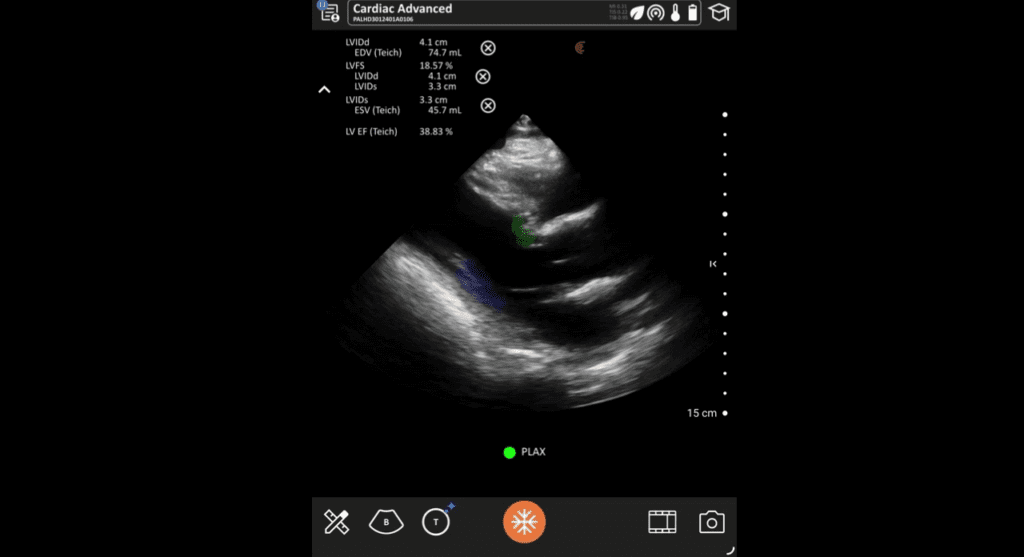
Clarius Ejection Fraction AI enables the physician to discover the answer in the room. The AI calculates left ventricular ejection fraction automatically from the cardiac scan, with no manual measurements or calipers required. What made it stand out for Dr. Guttman is where it calculates from. Many devices limit EF calculation to one or two views, typically the apical four-chamber or the parasternal long axis. Clarius EF AI works across multiple cardiac views.
What’s unique about the Clarius EF calculation is that it works across all the cardiac views. That adds real flexibility and practicality in a clinical setting.” — Dr. Joshua Guttman
In practice, this matters because not every patient gives you a perfect apical window. Obesity, COPD, body habitus, patient positioning, and time pressure mean clinicians often need to work with the best view available, not the ideal one. The ability to calculate EF from whichever view is accessible makes the feature meaningfully more useful in real clinical conditions.
For the primary care physician seeing a patient with unexplained fatigue or dyspnea, this is the difference between sending them away with a referral and no information and having an objective data point in the same visit that guides how urgently that referral needs to happen.
Clarius T-Mode Heart: Anatomy in Color, in Real Time
Cardiac ultrasound on a grayscale screen involves pattern recognition that develops over time. For clinicians who are earlier in their POCUS journey, that gray shimmer is one of the biggest barriers to building confidence with cardiac imaging. If you cannot tell what you are looking at, you stop trusting what you are seeing.
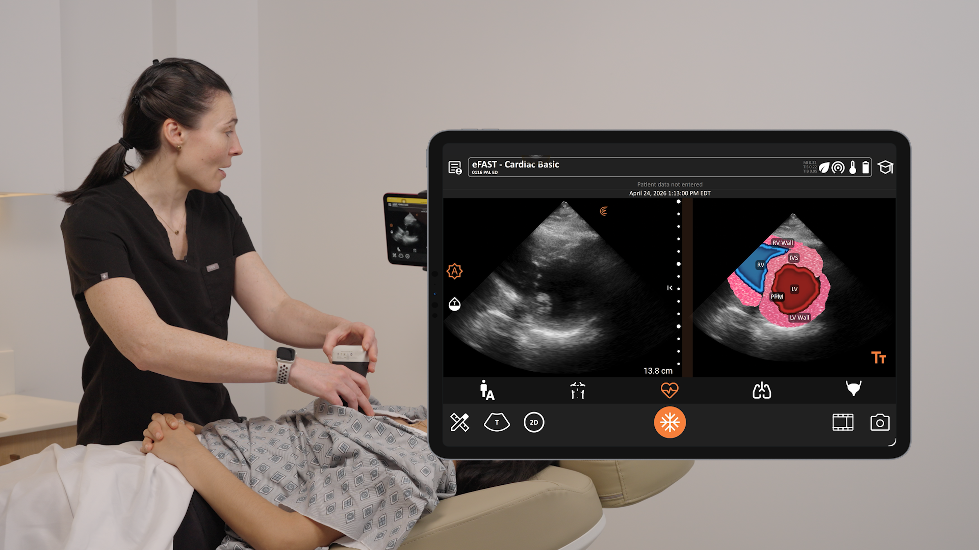
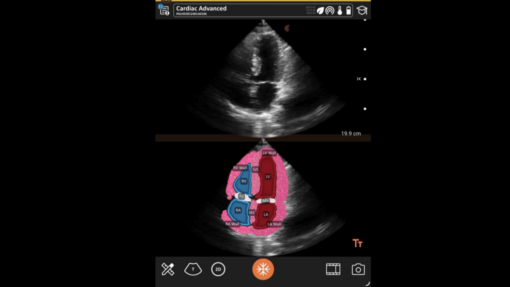
Clarius T-Mode changes that in real time. When Dr. Guttman activated T-Mode Heart during a parasternal long axis view, the result was immediate: a color-coded, labeled overlay appeared alongside the standard image. The RV was labeled in blue. The LV in red. Walls, valves, and structures were identified clearly, adjusting dynamically as he moved the probe.
Now go into the T-Mode and look how cool this is. It overlays with color exactly what I’m looking at. And even if I move, it adjusts. That’s really cool.” — Dr. Joshua Guttman
Dr. Guttman demonstrates T-Mode Heart in the parasternal long axis, short axis, apical four-chamber, and subxiphoid views. He tested it on pathology too. In a patient with dilated cardiomyopathy, T-Mode continued to label anatomy clearly even with an enlarged, poorly contracting LV. The small aortic valve was labeled. The relationship between chambers was visible in color even when the gray-scale image alone might have taken a less experienced eye longer to interpret.
He also notes that T-Mode images can be saved with the overlay intact, which makes them useful for documentation, teaching, and follow-up.
Built for the Learner, Useful for the Experienced Clinician
Dr. Guttman was direct about who benefits most from T-Mode: clinicians who are still developing their cardiac imaging confidence. The feature gives them an immediate visual check on what they are scanning.
But he also found it useful for verification in his own practice. When anatomy was ambiguous in grayscale, T-Mode provided a second layer of confirmation. For a primary care physician adding cardiac POCUS to their practice for the first time, that kind of real-time reinforcement is not a nice-to-have. It is what makes continued adoption realistic.
Clarius T-Mode Knee: Seeing What Other Devices Leave Gray
Dr. Guttman was unambiguous about where the Clarius PAL performs best: musculoskeletal imaging. And for the knee specifically, T-Mode Knee raises the bar.
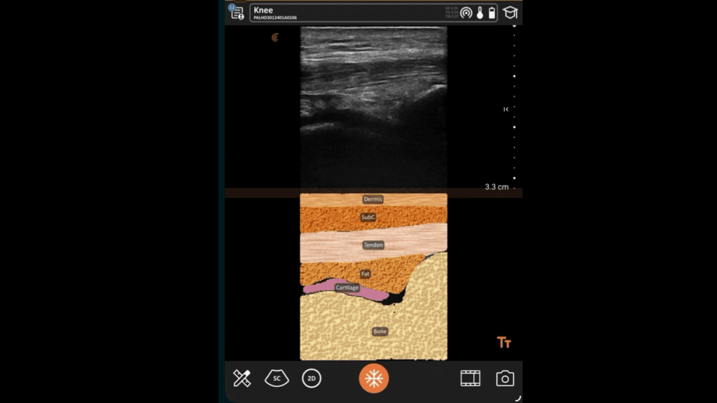
Scanning the anterior knee, Dr. Guttman pulls up the patella and the quad tendon clearly in grayscale. When he activates T-Mode, the image transforms: cartilage, fat, subcutaneous tissue, and bone each appear in distinct colors, labeled automatically, updating as he moves the probe across the anatomy.
I haven’t seen any other ultrasound that can do this, that tells you exactly what you’re looking at. You can see cartilage, fat, subcutaneous tissue, bone. Exactly what you’re looking at. And as you slide over, it follows the tendon and moves with it. This is ridiculously cool.” — Dr. Joshua Guttman
He demonstrates the patellar tendon down to its bony insertion, with T-Mode highlighting the insertion point. Then he moved to the Achilles, noting that T-Mode is not available there but that the grayscale tendon imaging was excellent regardless.
T-Mode Shoulder Adds Another Dimension
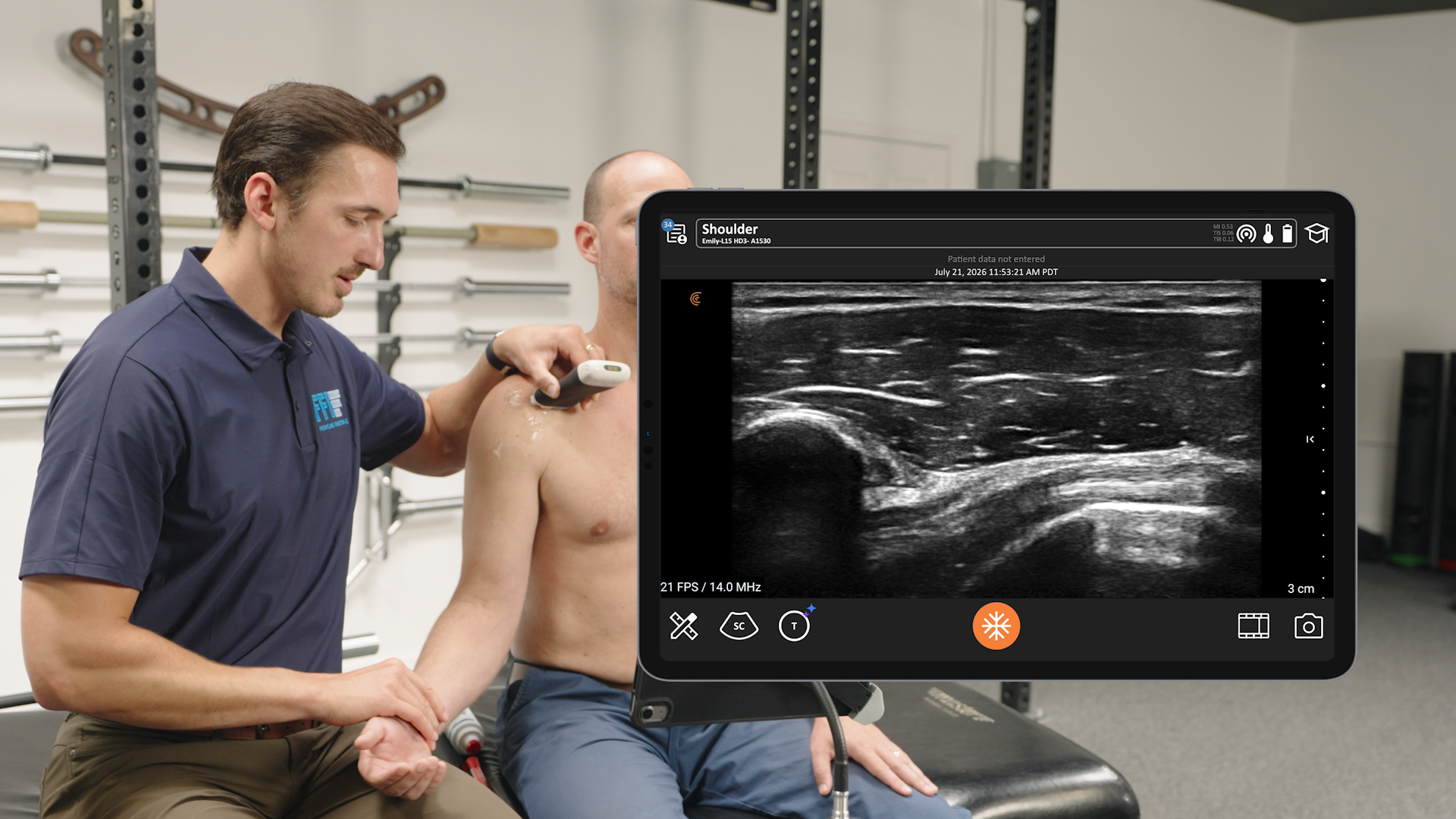
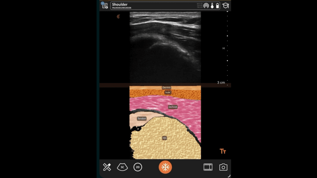
T-Mode is also available on the shoulder. Dr. Guttman scanned his own supraspinatus and noted that T-Mode Shoulder correctly identified the muscle and surrounding structures, adjusting the overlay as he changed his position. He points out that with a rotator cuff rupture, the structural discontinuity would be visible in both the grayscale and the color overlay.
Shoulder pain is a common complaint in primary care settings. T-Mode Shoulder gives clinicians a tool to learn ultrasound anatomy of the rotator cuff, so they can eventually become more skilled at recognizing pathology and managing patients more efficiently.
Dr. Guttman notes this is an area where Clarius has clearly invested, and where it delivers a different category of diagnostic confidence compared to standard handheld devices.
Clarius Bladder AI: A Volume Number That Changes the Visit
Bladder assessment in primary care often comes down to a clinical estimate. The patient has symptoms, the physical exam is inconclusive, and the options are limited: refer to urology, send to the ED, or manage empirically with incomplete information.
Clarius Bladder AI gives clinicians a third option: an objective bladder volume measurement in the exam room, in under a minute.
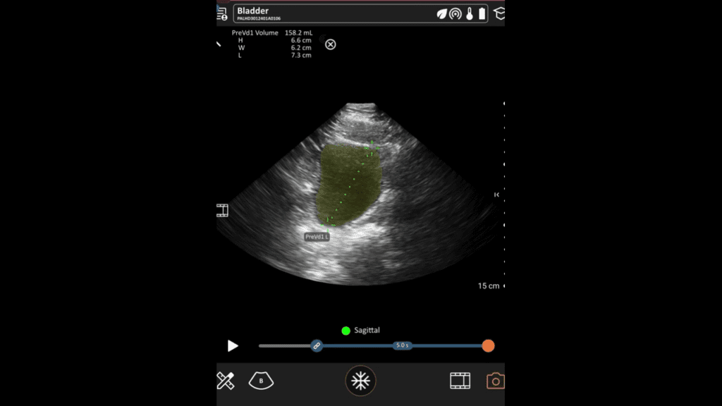
Dr. Guttman walks through the full workflow on screen. Scanning a well-filled bladder in both long axis and short axis, he navigates to the AI measurement tool, which automatically captures the bladder in the transverse view and produces dimensions on freeze. Moving to the sagittal view, the AI calculates and displays total volume on screen.
You go to measure, you can get it done with AI. It captures the whole bladder. Freeze, and it gives you the measurements. Then you unfreeze, go to the sagittal view, freeze again, and it gives you the volume. Pretty neat.” — Dr. Joshua Guttman
He also demonstrates the manual measurement option, showing that clinicians can perform the calculation themselves when preferred, with the AI workflow available as a faster alternative.
A Clinical Case Study of Urinary Retention
Dr. Guttman showed a clinical case of urinary retention: a clearly distended bladder with a thickened wall, and the prostate coming into view as the identified cause of retention. He noted that even without measuring, the volume was visually obvious. With measurement, the clinical decision becomes straightforward.
Pre-void and post-void residual measurements are both available in the workflow, making it useful for both retention assessment and post-void residual evaluation. For primary care physicians managing older patients with urinary symptoms, prostate conditions, or neurogenic bladder concerns, this is a capability that previously required referral to get.
The ability to document the volume, confirm the cause, and make the catheterization decision with confidence rather than guesswork is exactly the kind of outcome-level shift that Clarius Bladder AI is designed to produce.
Beyond the Four Features: A Full Clinical Picture
Dr. Guttman’s review covered more than the four highlighted features. In the full session, he scanned across a wide range of clinical applications, all on a single device.
In the abdomen, he demonstrated FAST exam views, gallbladder assessment, and identified a case of acute cholecystitis on POCUS that went directly to the operating room. He showed a small bowel obstruction with dilated loops measuring 4.5cm (normal under 2.5cm), identifying secondary signs including to-and-fro peristalsis.
In vascular imaging, he captured the carotid, internal jugular, and a clear view of the median nerve for nerve block guidance. He demonstrated a posterior tibial nerve block for a plantar laceration, with the needle visible in long axis alongside the target nerve.
On lung ultrasound, he identified B-lines in a patient with pulmonary edema secondary to the same systolic dysfunction seen in the cardiac views. The full clinical picture, from the cardiac dysfunction to the pulmonary consequence, visible on a single handheld device.
Dr. Guttman’s Conclusion about the Clarius PAL
Dr. Guttman’s independent review of the Clarius PAL HD3 comes down to this: it is a well-designed, full-capability device that earns its place in clinical practice, and that stands apart specifically because of its AI features.
The Clarius PAL HD3 is a really strong entry into the handheld ultrasound space. It’s a device that leans into being both a clinical and educational tool, with new AI features that make it genuinely useful for clinicians at any experience level.” — Dr. Joshua Guttman
About Dr. Joshua Guttman
Dr. Joshua Guttman is an emergency medicine physician and founder of Peachtree POCUS, a POCUS education and consulting firm. He has delivered more than 100 keynote addresses and workshops across North America, contributed to Rosen’s Emergency Medicine, and built a global audience through the Peachtree POCUS YouTube channel. He consults for leading medtech organizations and trains clinicians across medical specialties. Learn more at joshuaguttmanmd.com
Note: This review was sponsored by Clarius. Dr. Guttman retained full editorial independence. Clarius had no control over the content or opinions expressed in his review.
Make Ultrasound a Primary Advantage of Your Practice
Clarius brings high-definition imaging and smart tools right to the bedside, helping you make confident decisions and treat without leaving the exam room. If you’re interested in learning more about Clarius ultrasound for your exam room, a Clarius expert is available to provide a custom virtual demonstration.