Congestive heart failure (CHF) affects millions of patients annually and remains a leading cause of hospitalization. During this Clarius webinar, emergency physician and POCUS educator Dr. Tatiana Havryliuk illustrates how incorporating a structured, bedside point-of-care ultrasound (POCUS) protocol can instantly elevate clinical accuracy, optimize volume status management, and safely divert unnecessary hospital admissions.
To learn Dr. Havryliuk’s practical framework and watch ultrasound demonstrations, view the free webinar here: POCUS for Primary Care: Ultrasound Tips for Diagnosing and Managing CHF.
Read on for some highlights of Dr. Havryliuk’s presentation.
Using POCUS for Objective Clarity and Avoiding Diagnostic Pitfalls
Clinical impressions without imaging can inadvertently lock providers into an inaccurate diagnostic path. Janaye Smith, a sonographer at Clarius, highlighted two critical studies demonstrating how POCUS injects objective clarity into complex cardiopulmonary presentations:
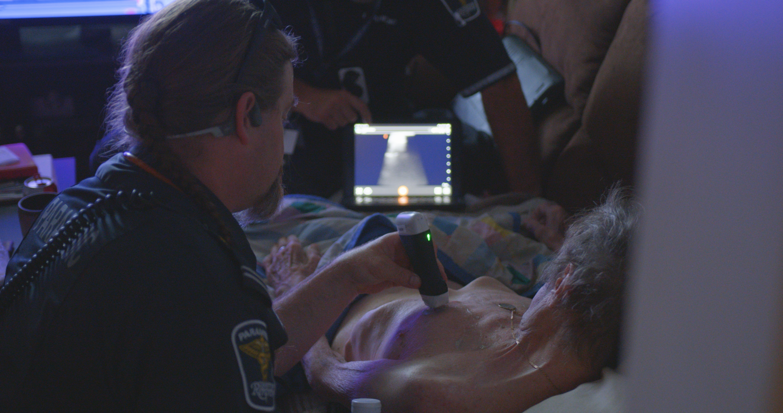
Pre-Hospital Accuracy: A study evaluating paramedics trained in lung POCUS showed they could reliably identify bilateral B-lines to diagnose pulmonary edema. This data demonstrates that minimal, structured training allows field providers to capture high-quality diagnostic windows that highly align with expert assessments. Read more about the field deployment of this modality in the Cambridge Core Review on Out-of-Hospital POCUS.
Mitigating Anchoring Bias: When “CHF” was noted in triage documentation, physicians were significantly less likely to investigate alternative causes of dyspnea, such as a pulmonary embolism. This clinical pattern was analyzed in a trial of over 108,000 patients, which you can review via the UCLA Health / JAMA Internal Medicine Research Release.
Integrating POCUS provides an objective, real-time examination of the heart, lungs, and inferior vena cava (IVC) to break through initial diagnostic assumptions and improve patient outcomes.
The 3-Step CHF POCUS Protocol
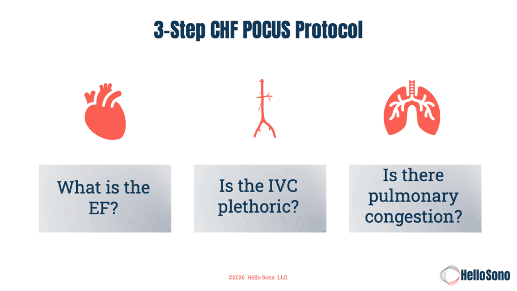
To keep things efficient for tight clinic schedules, Dr. Havryliuk recommends a simplified, three-step protocol designed to answer three distinct clinical questions.
1. Cardiac Scan ————> What is the approximate EF?
2. IVC Evaluation ———-> Is the IVC plethoric?
3. Lung Ultrasound ——-> Is there pulmonary congestion?
Video Demonstration: POCUS Assessment for CHF
Watch this 10-minute video; Dr. Havryliuk demonstrates how to scan key anatomy, including lung for pulmonary edema, the IVC for volume status, and focused echo for cardiac function for CHF assessment.
Step 1: The Cardiac Scan (Ejection Fraction)
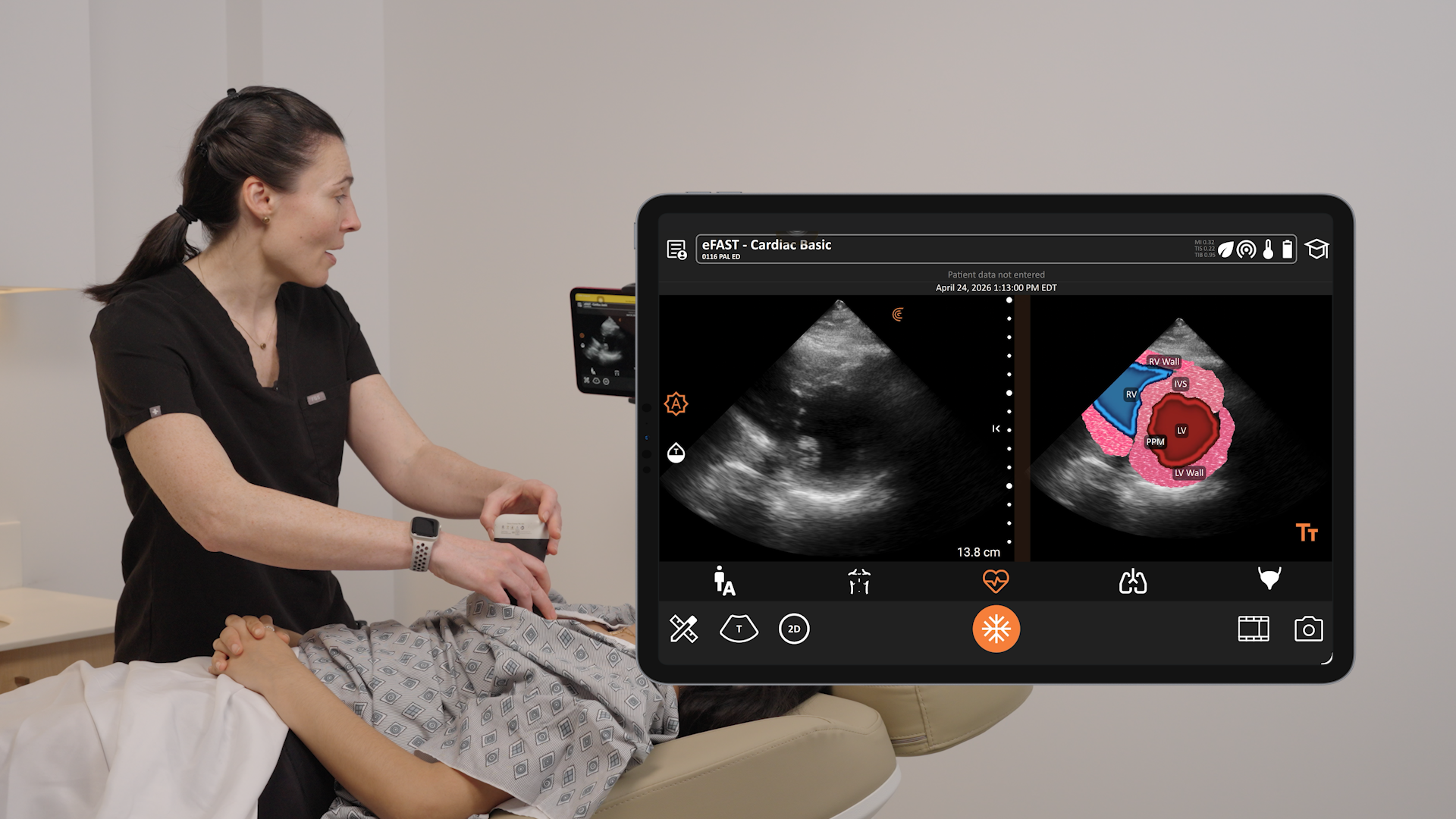
The goal is primarily to evaluate systolic function and approximate the left ventricular ejection fraction (EF). Dr. Havryliuk likes to evaluate LV function with a clockwise technique, starting with the parasternal window, followed by the apical, then subxipoid cardiac views.

- Parasternal Long Axis (PLAX) [10:00 Position]: Point the scanner marker toward the patient’s right shoulder. Ensure enough depth to see the descending aorta to differentiate pericardial from pleural effusions.
- Parasternal Short Axis (PSAX) [1:00 to 2:00 Position]: Rotate 90 degrees clockwise to achieve a circular cross-section of the left ventricle at the papillary muscle level.
- Apical Four-Chamber (A4C) [2:00 to 3:00 Position]: Slide to the apex. Dr. Havryliuk notes, “get your patient in a left [lateral decubitus] position for that apical four chamber view. It will make it so much easier to find.”
- Subcostal / Subxiphoid [6:00 Position]: Scan through the liver as your acoustic window to easily rule out pericardial effusions.
When estimating systolic function visually, look for three key markers: muscle thickening (the myocardium should thicken by roughly 30%), chamber volume change (LV volume should decrease by 50% or more during systole), and E-Point Septal Separation (EPSS). EPSS measures the distance between the anterior mitral valve leaflet tip and the interventricular septum.
Dr. Havryliuk’s Clinical Tip:
EPs is most useful. If it’s normal. Right. It’s a screening tool. If you got a normal EPs um then it is very unlikely that the ejection fraction is abnormal.”
EPSS Measurement Clinical Interpretation
< 7 mm Normal Ejection Fraction
7 mm – 13 mm Moderately Decreased Ejection Fraction
> 13 mm Severely Reduced Ejection Fraction
Step 2: Is the IVC Plethoric?
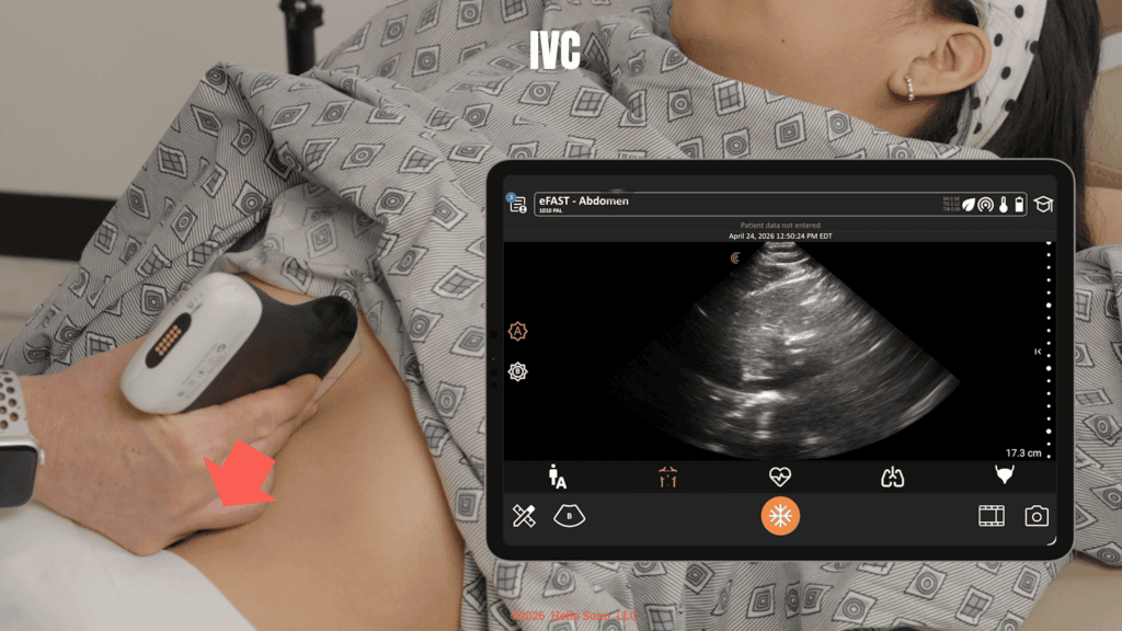
Move the scanner to the 6:00 position to trace the IVC where it empties into the right atrium, scanning through the liver. Evaluate its diameter and collapse ability during regular respiration approximately 2 cm distal to the right atrium.
- The Extremes Rule: Dr. Havryliuk emphasizes that IVC evaluation is most useful at its clinical extremes.
- Fluid Overload Status: An IVC that is dilated (greater than 2 cm) and shows less than 30% collapse during respiration is considered plethoric, strongly aligning with fluid overload.
- Fluid Responsiveness: Conversely, a highly collapsible IVC indicates the patient is highly likely to be fluid-responsive and is not in a systemic fluid overload state.
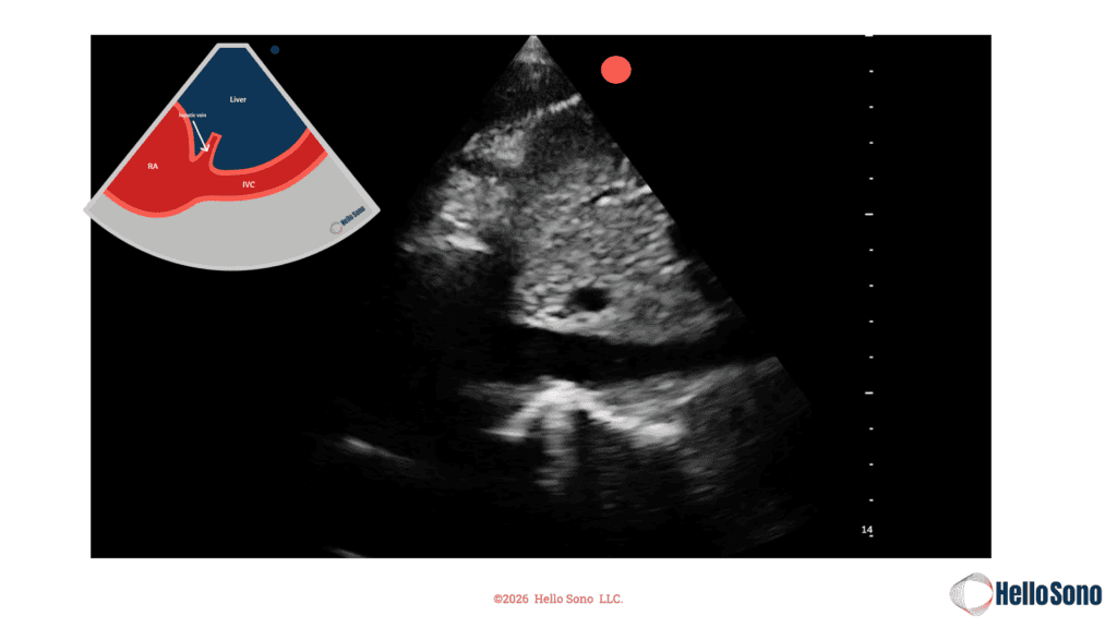
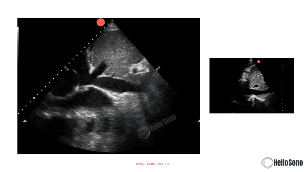
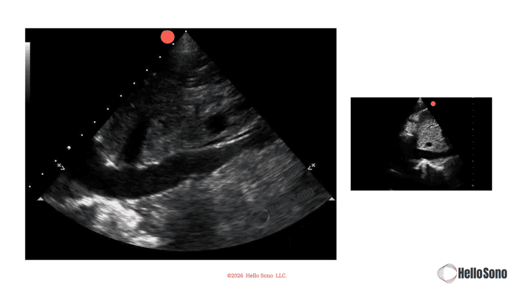
Step 3: Is There Pulmonary Congestion?
Scan at least three zones per lung (six fields total), including the apex, the posterior-lateral chest wall, and the lung bases. Because air reflects and scatters ultrasound waves, lung POCUS relies heavily on interpreting artifacts:
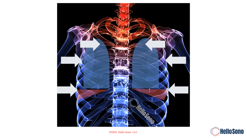
- A-Lines (Normal): Equidistant, horizontal reverberation lines indicating a normal, air-filled lung.
- B-Lines (Abnormal / Congestion): Vertical, laser-like “comet tails” arising from the pleura and extending to the bottom of the screen, indicative of fluid within the alveoli. Dr. Havryliuk notes that “in a patient with CHF exacerbation and fluid overload you should be seeing bilateral lines”.
- Spine Sign (Pleural Effusion): At the lung bases, fluid replaces air, allowing the ultrasound beam to visualize the thoracic spine continuing above the diaphragm.
Video Demonstration: Lung Ultrasound in a Primary Care Setting
In this detailed video, Dr. Havryliuk describes indications for lung ultrasound and the benefits of lung ultrasound in a primary care setting.
Real-World Case Applications
Dr. Havryliuk presents two contrasting patient scenarios that underscore how POCUS dramatically alters clinical pathways:
Case 1: The Standard Flare
A 72-year-old male with a history of CHF presented with mild dyspnea, leg swelling, and a history of missing his diuretics for two days.
- POCUS Findings: His EF was stable at ~35%, the IVC showed mild congestion, and bilateral B-lines were confined strictly to his lung bases.
- Outcome: Instead of sending him to the emergency department or waiting on laboratory panels, POCUS provided the safety data needed to confidently adjust his oral diuretics for outpatient management.
Case 2: The Diagnostic Masquerader
A 52-year-old female with a history of CHF presented with an almost identical story: dyspnea, leg swelling, and coarse breath sounds.
- POCUS Findings: Lung scanning revealed normal A-lines (no pulmonary congestion). However, a short-axis cardiac scan revealed a distinct “D-shaped” left ventricle caused by an enlarged right ventricle, paired with a non-collapsible, plethoric IVC.
- Outcome: The structural changes indicated severe right-sided heart strain. POCUS immediately shifted the working diagnosis from a heart failure flare to a life-threatening pulmonary embolism (PE), routing her straight to emergency care for anticoagulation.
Clinical Impact and Financial ROI of Incorporating POCUS in Primary Care Practice
Dr. Havryliuk showcases the measurable benefits of incorporating POCUS, including clinical performance, systemic cost reduction, and practice economics
Enhanced Diagnostic Performance of Using POCUS for CHF
A 2022 meta-analysis highlighted that ultrasound boasts a 92% sensitivity for detecting pulmonary congestion, starkly outperforming standard chest X-rays which sit at just 77% sensitivity. Review the comprehensive comparative data pooled across multiple international clinical cohorts via the PubMed Systematic Review and Meta-Analysis.
Furthermore, a study in primary care settings revealed that adding POCUS boosted diagnostic specificity for heart failure to 88%, compared to a poor 38% specificity when clinicians relied on clinical examination alone. Read the detailed methods, training parameters, and cardiologist-validated metrics in the PubMed Abstract on Primary Care POCUS for Heart Failure.
Financial Return on Investment (ROI) of Using POCUS in the United States
Whether operating under value-based care models or traditional fee-for-service frameworks, the financial incentives are distinct:
- Value-Based Care: A single CHF hospital admission costs roughly $15,000. Dr. Havryliuk notes that “preventing just one admission a month… is an opportunity to save $180,000 annually.”
- Fee-For-Service: Using ultrasound CPT codes for many procedures commands a conservative average global reimbursement of $180 per exam. Performing just one protocol per day opens up an estimated $42,000 in billable revenue annually.
Expert Q&A: Audience Questions Answered by Dr. Havryliuk
Q: Is JVD more reliable and accessible in a primary care setting than IVC? In a ten-minute appointment, it offers easier access to the neck without laying flat or assessing the abdomen.
Dr. Tatiana Havryliuk: “I would say definitely easier accessible. In terms of the evidence, there’s still more evidence for using IVC. Also, remember you are doing already cardiac and lung for the cardiac. You’re probably going to have the patients supine, so that wouldn’t be that much of an effort to get the IVC, especially if you’re already getting that subxiphoid view. It’s literally you’re turning the probe to get that IVC in the view. So, I think of it as not necessarily replacement unless you can’t get a good IVC view…”
Q: Do you always see bilateral B lines in fluid overload?
Dr. Tatiana Havryliuk: “You know, I don’t know the exact answer to this in my experience, yes. And the literature I’ve seen… in a patient with CHF exacerbation and fluid overload you should be seeing bilateral lines in someone who, for example has a focal pneumonia that’s forming, or maybe some scarring of the lung. You might be seeing some focal lines just on one side and one field.”
Q: Is CHF on ultrasound a late sign compared to stethoscope findings or chest X-rays?
Dr. Tatiana Havryliuk: “No. Early sign. See, ultrasound is really great with getting to these people, getting some confirmation and additional information early on in the course of their CHF.”
Q: Why is the IVC non-collapsible (plethoric) during a pulmonary embolism if it isn’t caused by fluid volume overload?
Dr. Tatiana Havryliuk: “When you see IVC that’s not collapsible, it could be either volume overload or pressure overload. The reason why IVC is not collapsible in a PE is their sudden change in pressure. There’s too much pressure on the right side of the heart.
Why Clarius Ultrasound is the Optimal Bedside Choice
During the webinar, Dr. Havryliuk and Janaye Smith showcased how the design of Clarius HD3 scanners addresses common barriers to POCUS clinical adoption:
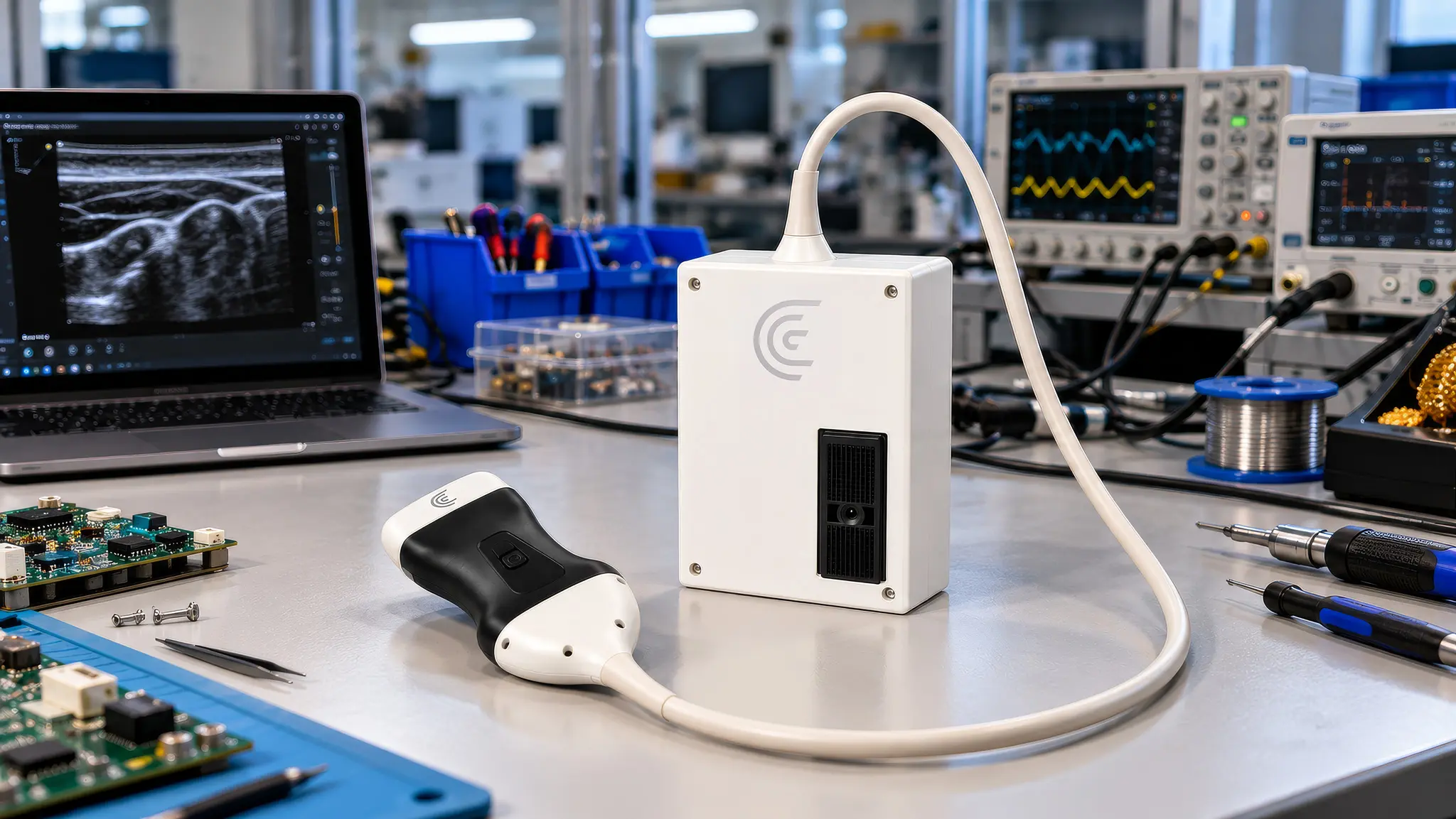
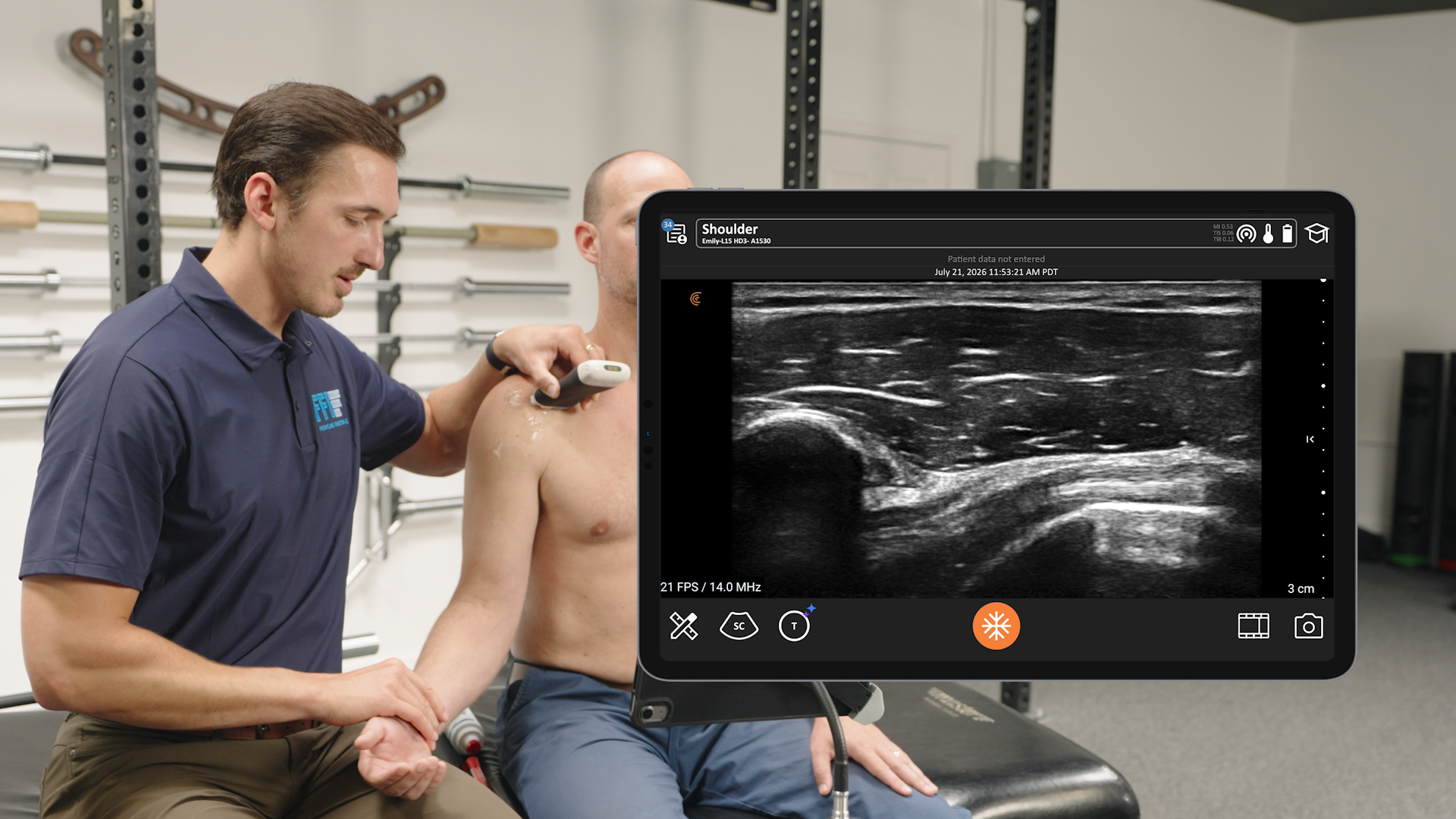
Clarius PAL Dual Array: This innovative scanner integrates both a phased array and a linear transducer in a single scanner. Clinicians can instantly switch from superficial tracking to deep cardiac, IVC, or lung configurations with a single tap in the app.
True Portability: The wireless design removes cords completely, maximizing mobility in tight exam spaces and enabling seamless sanitization, disinfection, or sterile sheathing.
AI-Assisted Workflows: Features like Auto EF and T-Mode take the guesswork out of image interpretation. The automated software tracks and computes ejection fractions directly on iOS or Android screens, offering immediate diagnostic verification for novice and expert users alike.
Learn more about Clarius POCUS for primary care. Feel free to schedule a private virtual demonstration at your convenience.
About Dr. Tatiana Havryliuk, M.D
Dr. Tatiana Havryliuk is an emergency physician, formerly ultrasound director at the Brooklyn Hospital, and founder of Hello Sono. Throughout her 15 years of clinical practice, she has relied on POCUS to make informed clinical decisions, whether it was in an urban ED, urgent care, or Everest Base Camp. Dr. Havryliuk is now on a mission to extend the benefits of POCUS to primary care and urgent care practices by addressing the key barriers: lack of POCUS competency and operational readiness. She and her team at Hello Sono offer in-person provider training and support with credentialing and implementation to build high-quality, compliant, and financially sound POCUS programs.