Clinical skin evaluation in aesthetic medicine has historically relied on surface observations, photography, and manual palpation. While these methods serve a foundational purpose, they carry inherent limitations. Photographic outcomes are highly susceptible to lighting biases and angle variations, while palpation remains subjective and clinician-dependent. Furthermore, patient self-assessment is frequently driven by emotional fluctuations rather than measurable physiological changes.
As the field shifts toward regenerative medicine, diagnostic and evaluative tools must evolve accordingly. Aesthetic practices are transitioning away from purely visual impressions and toward a quantifiable, objective framework.
During a recent Clarius webinar, Ultrasound for Facial Aesthetics: Skin Evaluation and Biostimulation Assessment, aesthetics physician Dr. Gina Moret and Clarius sonographer Janaye Smith demonstrated how high-frequency point-of-care ultrasound (POCUS) provides this missing link. Acting as a real-time, non-invasive tool for the skin, ultrasound allows clinicians to look past surface aesthetics and directly analyze tissue-level data to prove exactly what is happening beneath the surface.

The future of aesthetic medicine is not about performing treatments. It is about understanding the tissue, measuring the change and providing natural results.” — Dr. Gina Moret
Establishing the Measurement Baseline: Normal vs. Aged Tissue Architectures
Before accurately interpreting structural changes induced by aesthetic treatments, clinicians must understand normal tissue architecture under high-frequency ultrasound. A standard cutaneous scan reveals a highly echogenic, thin surface line representing the epidermis. Directly beneath lies the dermis, which presents a brighter echogenic pattern due to its dense collagen composition. Below the dermal-hypodermal junction, the hypodermis is characterized by dark, hypoechoic fat globules interspersed with thin lines of fibrotic septae.
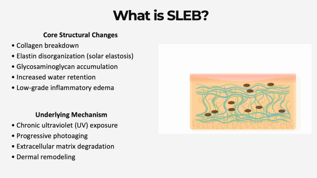
When evaluating aging or photo-damaged skin, a distinct pathological marker appears: the SLEB (Sub-epidermal Low-echogenic Band).
- Pathophysiology: The SLEB presents as a dark, hypoechoic horizontal band located directly underneath the epidermis.
- Structural Biomarker: It serves as a direct ultrasound marker of advanced skin aging, chronic UV exposure, and oxidative damage.
- Histological Characteristics: This low-echogenic zone is driven by localized extracellular matrix breakdown, water retention from low-grade chronic inflammation, and solar elastosis. This refers to the pathological accumulation and degeneration of abnormal elastin fibers replacing organized collagen architecture.
- Morphological Irregularity: The SLEB is highly variable in thickness and features an irregular distribution across the treatment area, especially in severely photo-aged tissue.
The Calculation Metric: Quantifying Dermal Degradation via the SLEB Ratio Framework
Dr. Moret emphasizes that absolute SLEB thickness cannot be interpreted in isolation. A 0.2 mm SLEB within a naturally thick dermal layer may indicate minor alteration, whereas that same 0.2 mm measurement within a thin, atrophic dermal layer represents severe structural compromise. To achieve a standardized parameter independent of anatomical site or natural skin variations, clinicians must calculate the SLEB Ratio:
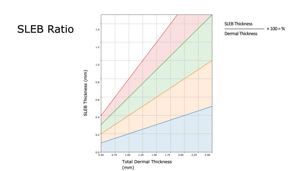
When performing this calculation, it is clinically vital that the total dermal thickness measurement completely excludes the epidermis but includes the full thickness of the SLEB layer down to the upper border of the hypodermis. The resulting percentage reflects exactly how much of the healthy dermal compartment has been structurally altered or displaced by degenerative tissue changes.
Predictive Treatment Mapping: Matching Biostimulators to Objective Dermal Metrics
Quantifying the SLEB ratio allows clinicians to shift away from generalized treatment choices and design highly targeted, customized bio stimulation protocols to maximize cellular response:
- 0% to 10% (Optimal Skin Structure): The SLEB is minimal or entirely absent, accompanied by a bright, highly homogeneous dermis. Clinical strategy focuses on preservation using non-aggressive maintenance protocols, such as one to two cycles of mesotherapy or skin boosters annually.
- 10% to 20% (Early Structural Changes): A thin SLEB is identifiable, though robust background dermal echogenicity remains intact. This zone calls for early, targeted interventions to halt matrix degradation, utilizing Platelet-Rich Plasma (PRP), PDRN (polynucleotides/salmon DNA), or light bio stimulative therapies.
- 20% to 30% (Moderate Dermal Aging): The tissue presents early dermal heterogeneity and noticeably reduced brightness. Active regeneration is required to restore dermal architecture, utilizing focused polynucleotide regimens, exosomes, and combination tightening therapies.
- 30% to 40% (Advanced Dermal Compromise): Marked by a significantly thicker SLEB and a dark, disorganized dermis indicating widespread collagen loss. Stronger therapeutic interventions must be deployed, specifically particulate biostimulators like calcium hydroxyapatite (CaHA) or poly-L-lactic acid / poly-D,L-lactic acid (PLLA/PDLLA).
- Over 40% (Severe Photo-Damage): Characterized by severe dermal thinning, a highly dense and thickened SLEB, and loss of structural definition. These fragile tissues have a slowed regenerative capacity and require a cautious, multi-modal approach combining high-intensity focused ultrasound (HIFU), deep biostimulators, and advanced regenerative therapies.
VIDEO Demonstration: SLEB Measurements to Predict Skin Aging
Watch this 4-minute video to see Dr. Moret precisely demonstrate how to use the Clarius L20 HD3 for the measurement of the SLEB (subepidermal low echogenic band) and the dermis to help predict skin aging.
Documenting Results over Time: Measuring Tissue Responses and Cellular Remodeling
Using high-frequency ultrasound allows practitioners to verify whether true dermal remodeling and cellular regeneration actually occur post-treatment. Rather than relying on subjective observation, clinicians can track distinct product behaviors and indirect tissue healing signatures over time:
Calcium Hydroxyapatite (CaHA): Immediately post-injection, the product appears as highly visible, hyper-echogenic particulate deposits. Over a two-month timeline, successful tissue integration and neo-collagenesis are documented by the emergence of a distinct, thick hyper-echogenic band, reflecting increased structural density.
PLLA and PDLLA Compounds: These biostimulators display subtle localized product deposits characterized by a telltale posterior acoustic shadow on ultrasound. At one month, the surrounding tissue demonstrates progressive, uniform dermal and subdermal echogenic maturation.
High-Intensity Focused Ultrasound (HIFU): Ultrasound allows clinicians to confirm that thermal energy is safely delivered to the intended target layer, such as the SMAS, preventing inadvertent fat injury. Successful remodeling reveals extra hyper-dense focal “thermal dots” and a distinctly brighter, consolidated SMAS layer.
Exosomes and Liquid Regenerative Therapies: While liquid particles cannot be directly visualized after injection, ultrasound tracks their indirect regenerative efficacy. Over time, clinicians look for quantified reductions in the SLEB ratio, improved tissue homogeneity, and a measurable increase in total dermal thickness. Color Doppler mode is highly effective here to document the neo-vascularization essential for long-term tissue health.
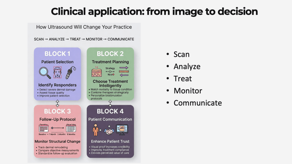
To maintain clinical accuracy, evaluations must follow a strict, highly standardized follow-up loop. Dr. Moret advises scanning patients at baseline (pre-treatment), 1 month (Early tissue integration), 3 months (Peak collagen response), and 6 months (log-term remodelling) post-procedure using identical scanning parameters, positioning, and anatomical landmarks.
Optimizing the Workflow: How Advanced POCUS Flattens the Learning Curve
Transitioning from a blind injection methodology to a POCUS-guided practice requires an ultra-high-frequency tool that eliminates technical barriers. Dr. Moret and the Clarius team highlighted several ways the Clarius L20 HD3 shortens this learning curve to make quantification a seamless part of your workflow:
Real-Time T-Mode Artificial Intelligence: Learning facial ultrasound patterns takes time, but the integrated T-Mode for Facial Aesthetics feature accelerates proficiency. The AI algorithm automatically colors and labels distinct tissue layers—such as the SMAS, muscles, and deep fascia—side by side with the grayscale image, providing immediate diagnostic clarity. As Dr. Moret noted, “The artificial intelligence is going to show you all of the tissues that are there in color… that makes learning very fast.” Watch the video demonstration:
Ergonomics and Portability: The wireless design frees up physical space in the clinical room and allows for swift disinfection between patients. The scanner is compact and accessible enough to easily incorporate into daily workflows. Dr. Moret shared, “When I first got my L20, I used it as a toy. I scanned my dog, I scanned my kids because it’s so much fun… You can carry it with you, it’s amazing.”
Visual Proof for Patient Communication: Sharing the scanning screen live with patients shifts the clinical dynamic. When patients visually understand their underlying tissue quality and actively see their structural SLEB ratio improve, it builds clinical credibility, deepens patient trust, and significantly enhances compliance for multi-session regenerative protocols.
Expert Insights: Webinar Audience Q&A
During the live broadcast, Dr. Moret addressed several highly relevant clinical questions regarding standardizing and isolating skin measurements:
Q: Are there specific systemic pathologies or medications that can actively alter or skew baseline SLEB measurements? Dr. Gina Moret: Yes, there are. Chronic corticosteroid use induces pronounced skin atrophy, which thins the epidermis and artificially inflates the relative proportion of the SLEB within the dermal layer. Systemic connective tissue disorders—such as scleroderma, cutaneous lupus, and psoriasis—directly degrade the extracellular matrix and drive severe low-grade inflammation, altering the baseline architecture. Additionally, in my personal clinical experience, I have noted structural variations in patients undergoing active isotretinoin therapy. If a patient presents a SLEB ratio exceeding 45% to 50%, you should immediately re-scan to rule out technical error, and thoroughly evaluate them for an underlying clinical dermatological condition.
Q: With the widespread rise of GLP-1 receptor agonist therapies, are we observing structural changes localized strictly to the facial fat pads, or is there a direct impact on the deeper dermal architecture? Dr. Gina Moret: The most prominent and immediate changes we observe on ultrasound are concentrated within the structural fat pads. However, anecdotally, I have also consistently observed a distinct thinning and loosening of the SMAS layer and the primary supporting fibrotic septae of the face. Gathering tightly standardized, peer-reviewed dermal data on GLP-1 patients remains challenging due to varying patient timelines and baseline variations. However, the tissue laxity visualized under ultrasound highlights that these patients require targeted structural remodeling that goes far beyond simple volume replacement.
Q: Should a clinician assume that a SLEB measurement taken at a single anatomical site is representative of the entire face? Dr. Gina Moret: Definitely not; they do differ from different areas. That is why you have to standardize the specific areas that you are going to check. In my practice, I systematically evaluate and track six specific zones: the zygomatic arch, the temples, the jawline, the infraorbital region (under eye), the perioral area (mouth), and the neck.
Watch Dr. Moret’s Webinar for Practical Tips on Advancing Your Aesthetics Practice
Moving from perception to precision ensures patient safety, maps crucial vascular zones, and provides undeniable verification of cellular regeneration. Watch the full webinar for Dr. Moret’s complete presentation, which is now available on-demand.
Interested in Implementing Dr. Moret’s Techniques at Your Practice?
Schedule a personalized virtual demo to learn more about Clarius. We’ll show you how easy it is to integrate handheld ultrasound into your workflow.
About Dr. Gina Moret
Doctor Gina Moret was born in Brazil and graduated from a medical university in Brazil at FESO University in 2008. She is a board-certified physician, expert in the advanced use of dermal fillers, botulinum toxin, and spider web technique, specializing in minimally invasive holistic aesthetic and regenerative medicine. She is known for her artistic and natural approach to aesthetics. She has pursued her training in aesthetic Medicine with some of the world’s experts in the field. Successfully completing many medical education extension programs in France, İtaly, Brazil, Singapore, and Turkey. Doctor Moret is a master trainer for Yvoire – LG fillers, Infinite Threads, and is the Head Trainer and Educational Director of B Academy. She is also the first and only doctor in South America to be legally accredited in the Republic of Turkey. Because Dr. Moret is a firm believer in education, she has successfully obtained international accreditation as a medical trainer and education supervisor in the United Kingdom.